1OM9

Structure of the GGA1-appendage in complex with the p56 binding peptide


Experimental Data Snapshot

  • Method: X-RAY DIFFRACTION
  • Resolution: 2.50 Å
  • R-Value Free: 0.262 
  • R-Value Work: 0.213 
  • R-Value Observed: 0.216 

wwPDB Validation   3D Report Full Report


This is version 1.3 of the entry. See complete history


Literature

Structural basis for binding of accessory proteins by the appendage domain of GGAs

Collins, B.M.Praefcke, G.J.K.Robinson, M.S.Owen, D.J.

(2003) Nat Struct Biol 10: 607-613

  • DOI: https://doi.org/10.1038/nsb955
  • Primary Citation of Related Structures:  
    1OM9

  • PubMed Abstract: 

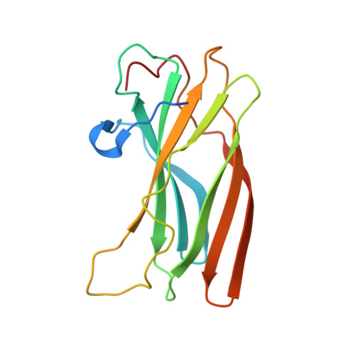
    The Golgi-associated, gamma-adaptin-related, ADP-ribosylation-factor binding proteins (GGAs) and adaptor protein (AP)-1 are adaptors involved in clathrin-mediated transport between the trans-Golgi network and endosomal system. The appendage domains of GGAs and the AP-1 gamma-adaptin subunit are structurally homologous and have been proposed to bind to accessory proteins via interaction with short sequences containing phenylalanines and acidic residues. Here we present the structure of the human GGA1 appendage in complex with its cognate binding peptide from the p56 accessory protein (DDDDFGGFEAAETFD) as determined by X-ray crystallography. The interaction is governed predominantly by packing of the first two phenylalanine residues of the peptide with conserved basic and hydrophobic residues from GGA1. Additionally, several main chain hydrogen bonds cause the peptide to form an additional beta-strand on the edge of the preexisting beta-sheet of the protein. Isothermal titration calorimetry was used to assess the affinities of different peptides for the GGA and gamma-appendage domains.


  • Organizational Affiliation

    Cambridge Institute for Medical Research, Department of Clinical Biochemistry, University of Cambridge, Hills Road, Cambridge CB2 2XY, UK. bmc25@cam.ac.uk


Macromolecules
Find similar proteins by:  (by identity cutoff)  |  3D Structure
Entity ID: 1
MoleculeChains Sequence LengthOrganismDetailsImage
ADP-ribosylation factor binding protein GGA1A,
C [auth B]
154Homo sapiensMutation(s): 0 
Gene Names: GGA1
Membrane Entity: Yes 
UniProt & NIH Common Fund Data Resources
Find proteins for Q9UJY5 (Homo sapiens)
Explore Q9UJY5 
Go to UniProtKB:  Q9UJY5
PHAROS:  Q9UJY5
GTEx:  ENSG00000100083 
Entity Groups  
Sequence Clusters30% Identity50% Identity70% Identity90% Identity95% Identity100% Identity
UniProt GroupQ9UJY5
Sequence Annotations
Expand
  • Reference Sequence

Find similar proteins by:  Sequence   |   3D Structure  

Entity ID: 2
MoleculeChains Sequence LengthOrganismDetailsImage
15-mer peptide fragment of p56B [auth P],
D [auth Q]
15N/AMutation(s): 0 
Membrane Entity: Yes 
UniProt & NIH Common Fund Data Resources
Find proteins for Q7Z6B0 (Homo sapiens)
Explore Q7Z6B0 
Go to UniProtKB:  Q7Z6B0
PHAROS:  Q7Z6B0
GTEx:  ENSG00000123106 
Entity Groups  
Sequence Clusters30% Identity50% Identity70% Identity90% Identity95% Identity100% Identity
UniProt GroupQ7Z6B0
Sequence Annotations
Expand
  • Reference Sequence
Experimental Data & Validation

Experimental Data

  • Method: X-RAY DIFFRACTION
  • Resolution: 2.50 Å
  • R-Value Free: 0.262 
  • R-Value Work: 0.213 
  • R-Value Observed: 0.216 
  • Space Group: P 31 2 1
Unit Cell:
Length ( Å )Angle ( ˚ )
a = 61.414α = 90
b = 61.414β = 90
c = 145.008γ = 120
Software Package:
Software NamePurpose
MOSFLMdata reduction
SCALAdata scaling
AMoREphasing
REFMACrefinement
CCP4data scaling

Structure Validation

View Full Validation Report


Entry History 

Deposition Data

Revision History  (Full details and data files)

  • Version 1.0: 2003-07-29
    Type: Initial release
  • Version 1.1: 2008-04-29
    Changes: Version format compliance
  • Version 1.2: 2011-07-13
    Changes: Version format compliance
  • Version 1.3: 2023-08-16
    Changes: Data collection, Database references, Refinement description