1OKC

structure of mitochondrial ADP/ATP carrier in complex with carboxyatractyloside


Experimental Data Snapshot

  • Method: X-RAY DIFFRACTION
  • Resolution: 2.20 Å
  • R-Value Free: 0.266 
  • R-Value Work: 0.220 
  • R-Value Observed: 0.220 

wwPDB Validation   3D Report Full Report


Ligand Structure Quality Assessment 


This is version 1.1 of the entry. See complete history


Literature

Structure of Mitochondrial Adp/ATP Carrier in Complex with Carboxyatractyloside

Pebay-Peyroula, E.Dahout-Gonzalez, C.Kahn, R.Trezeguet, V.Lauquin, G.J.-M.Brandolin, G.

(2003) Nature 426: 39

  • DOI: https://doi.org/10.1038/nature02056
  • Primary Citation of Related Structures:  
    1OKC

  • PubMed Abstract: 

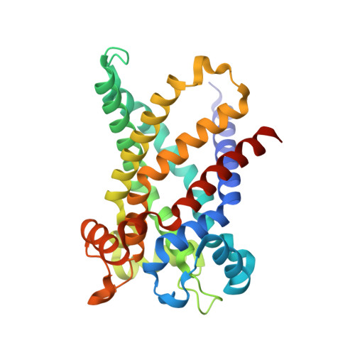
    ATP, the principal energy currency of the cell, fuels most biosynthetic reactions in the cytoplasm by its hydrolysis into ADP and inorganic phosphate. Because resynthesis of ATP occurs in the mitochondrial matrix, ATP is exported into the cytoplasm while ADP is imported into the matrix. The exchange is accomplished by a single protein, the ADP/ATP carrier. Here we have solved the bovine carrier structure at a resolution of 2.2 A by X-ray crystallography in complex with an inhibitor, carboxyatractyloside. Six alpha-helices form a compact transmembrane domain, which, at the surface towards the space between inner and outer mitochondrial membranes, reveals a deep depression. At its bottom, a hexapeptide carrying the signature of nucleotide carriers (RRRMMM) is located. Our structure, together with earlier biochemical results, suggests that transport substrates bind to the bottom of the cavity and that translocation results from a transient transition from a 'pit' to a 'channel' conformation.


  • Organizational Affiliation

    Institut de Biologie Structurale, UMR 5075 CEA-CNRS-Université Joseph Fourier, 41 rue Jules Horowitz, F-38027, Grenoble cedex 1, France. pebay@ibs.fr


Macromolecules
Find similar proteins by:  (by identity cutoff)  |  3D Structure
Entity ID: 1
MoleculeChains Sequence LengthOrganismDetailsImage
ADP, ATP CARRIER PROTEIN HEART ISOFORM T1297Bos taurusMutation(s): 0 
Membrane Entity: Yes 
UniProt
Find proteins for P02722 (Bos taurus)
Explore P02722 
Go to UniProtKB:  P02722
Entity Groups  
Sequence Clusters30% Identity50% Identity70% Identity90% Identity95% Identity100% Identity
UniProt GroupP02722
Sequence Annotations
Expand
  • Reference Sequence
Small Molecules
Ligands 4 Unique
IDChains Name / Formula / InChI Key2D Diagram3D Interactions
CDL
Query on CDL

Download Ideal Coordinates CCD File 
C [auth A],
D [auth A],
E [auth A]
CARDIOLIPIN
C81 H156 O17 P2
XVTUQDWPJJBEHJ-KZCWQMDCSA-L
PC1
Query on PC1

Download Ideal Coordinates CCD File 
H [auth A],
I [auth A],
J [auth A],
K [auth A]
1,2-DIACYL-SN-GLYCERO-3-PHOSPHOCHOLINE
C44 H88 N O8 P
NRJAVPSFFCBXDT-HUESYALOSA-N
CXT
Query on CXT

Download Ideal Coordinates CCD File 
B [auth A]Carboxyatractyloside
C31 H46 O18 S2
AQFATIOBERWBDY-LNQSNDDKSA-N
LDM
Query on LDM

Download Ideal Coordinates CCD File 
F [auth A],
G [auth A]
3-LAURYLAMIDO-N,N'-DIMETHYLPROPYLAMINOXYDE
C17 H36 N2 O2
JNGWKQJZIUZUPR-UHFFFAOYSA-N
Experimental Data & Validation

Experimental Data

  • Method: X-RAY DIFFRACTION
  • Resolution: 2.20 Å
  • R-Value Free: 0.266 
  • R-Value Work: 0.220 
  • R-Value Observed: 0.220 
  • Space Group: P 21 21 2
Unit Cell:
Length ( Å )Angle ( ˚ )
a = 85.437α = 90
b = 83.463β = 90
c = 49.922γ = 90
Software Package:
Software NamePurpose
CNSrefinement
DENZOdata reduction
CCP4data scaling
CCP4phasing

Structure Validation

View Full Validation Report


Ligand Structure Quality Assessment 


Entry History 

Deposition Data

Revision History  (Full details and data files)

  • Version 1.0: 2003-11-07
    Type: Initial release
  • Version 1.1: 2014-01-29
    Changes: Advisory, Derived calculations, Non-polymer description, Other, Refinement description, Source and taxonomy, Structure summary, Version format compliance