


Crystal Structure of Yhbo from Escherichia Coli
Claude, J.B., Abergel, C., Claverie, J.M.To be published.
Experimental Data Snapshot
wwPDB Validation 3D Report Full Report

Entity ID: 1 | |||||
|---|---|---|---|---|---|
| Molecule | Chains | Sequence Length | Organism | Details | Image |
| HYPOTHETICAL PROTEIN YHBO | 193 | Escherichia coli K-12 | Mutation(s): 0 | 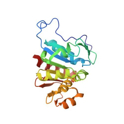 | |
UniProt | |||||
Find proteins for P45470 (Escherichia coli (strain K12)) Explore P45470 Go to UniProtKB: P45470 | |||||
Entity Groups | |||||
| Sequence Clusters | 30% Identity50% Identity70% Identity90% Identity95% Identity100% Identity | ||||
| UniProt Group | P45470 | ||||
Sequence AnnotationsExpand | |||||
| |||||
| Length ( Å ) | Angle ( ˚ ) |
|---|---|
| a = 68.037 | α = 90 |
| b = 72.446 | β = 90 |
| c = 75.946 | γ = 90 |
| Software Name | Purpose |
|---|---|
| CNS | refinement |
| MOSFLM | data reduction |
| SCALEPACK | data scaling |
| AMoRE | phasing |
RCSB PDB (citation) is hosted by
RCSB PDB is a member of the

