Structure of the Bifunctional Dctp Deaminase-Dutpase from Methanocaldococcus Jannaschii and its Relation to Other Homotrimeric Dutpases
Johansson, E., Bjornberg, O., Nyman, P.O., Larsen, S.(2003) J Biol Chem 278: 27916
- PubMed: 12756253
- DOI: https://doi.org/10.1074/jbc.M304361200
- Primary Citation of Related Structures:
1OGH - PubMed Abstract:
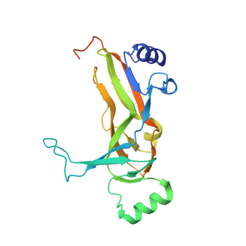
The bifunctional dCTP deaminase-dUTPase (DCD-DUT) from Methanocaldococcus jannaschii catalyzes the deamination of the cytosine moiety in dCTP and the hydrolysis of the triphosphate moiety forming dUMP, thereby preventing uracil from being incorporated into DNA. The crystal structure of DCD-DUT has been determined to 1.88-A resolution and represents the first known structure of an enzyme catalyzing dCTP deamination. The functional form of DCD-DUT is a homotrimer wherein the subunits are composed of a central distorted beta-barrel surrounded by two beta-sheets and four helices. The trimeric DCD-DUT shows structural similarity to trimeric dUTPases at the tertiary and quaternary levels. There are also additional structural elements in DCD-DUT compared with dUTPase because of a longer primary structure. Four of the five conserved sequence motifs that create the active sites in dUTPase are found in structurally equivalent positions in DCD-DUT. The last 25 C-terminal residues of the 204-residue-long DCD-DUT are not visible in the electron density map, but, analogous to dUTPases, the C terminus is probably ordered, closing the active site upon catalysis. Unlike other enzymes catalyzing the deamination of cytosine compounds, DCD-DUT is not exploiting an enzyme-bound metal ion such as zinc or iron for nucleophile generation. The active site contains two water molecules that are engaged in hydrogen bonds to the invariant residues Ser118, Arg122, Thr130, and Glu145. These water molecules are potential nucleophile candidates in the deamination reaction.
Organizational Affiliation:
Center for Crystallographic Studies, Department of Chemistry, University of Copenhagen, Universitetsparken 5, DK-2100 Copenhagen Ø, Denmark.