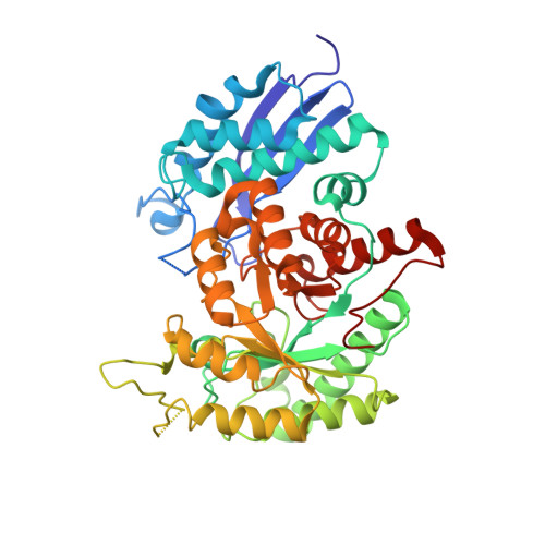
The Crystal Structure of Trypanosoma Brucei Enolase: Visualisation of the Inhibitory Metal Binding Site III and Potential as Target for Selective, Irreversible Inhibition
Da Silva Giotto, M.T., Hannaert, V., Vertommen, D., Navarro, M.V.A.S., Rider, M.H., Michels, P.A.M., Garratt, R.C., Rigden, D.J.(2003) J Mol Biol 331: 653
- PubMed: 12899835
- DOI: https://doi.org/10.1016/s0022-2836(03)00752-6
- Primary Citation of Related Structures:
1OEP - PubMed Abstract:
The glycolytic enzymes of the trypanosomatids, that cause a variety of medically and agriculturally important diseases, are validated targets for drug design. Design of species-specific inhibitors is facilitated by the availability of structural data. Irreversible inhibitors, that bound covalently to the parasite enzyme alone, would be potentially particularly effective. Here we determine the crystal structure of enolase from Trypanosoma brucei and show that two cysteine residues, located in a water-filled cavity near the active-site, are modified by iodoacetamide leading to loss of catalytic activity. Since these residues are specific to the Trypanosomatidae lineage, this finding opens the way for the development of parasite-specific, irreversibly binding enolase inhibitors. In the present structure, the catalytic site is partially occupied by sulphate and two zinc ions. Surprisingly, one of these zinc ions illustrates the existence of a novel enolase-binding site for divalent metals. Evidence suggests that this is the first direct visualization of the elusive inhibitory metal site, whose existence has hitherto only been inferred from kinetic data.
Organizational Affiliation:
Instituto de Física de São Carlos, Universidade de São Paulo, São Carlos SP, Brazil.