1OE4

Xenopus SMUG1, an anti-mutator uracil-DNA Glycosylase


Experimental Data Snapshot

  • Method: X-RAY DIFFRACTION
  • Resolution: 2.00 Å
  • R-Value Free: 0.216 
  • R-Value Work: 0.177 
  • R-Value Observed: 0.179 

wwPDB Validation   3D Report Full Report


This is version 1.1 of the entry. See complete history


Literature

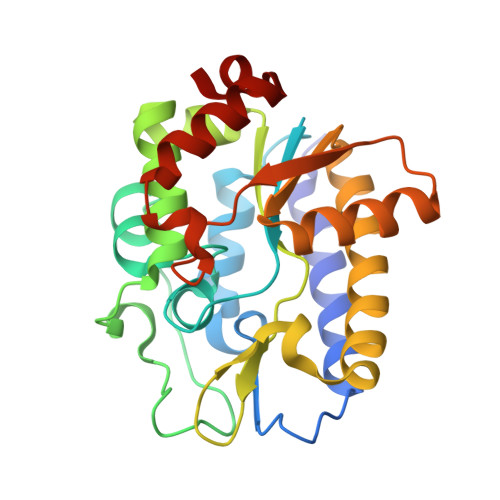
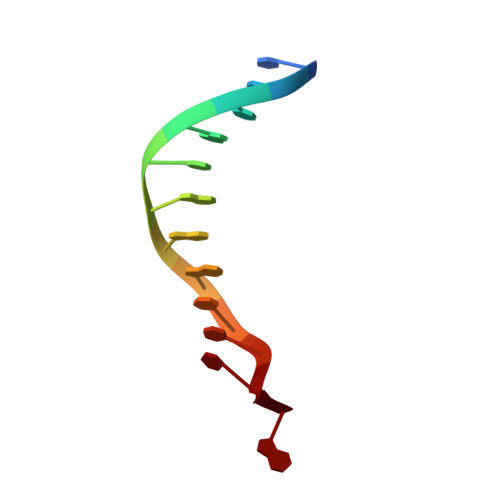
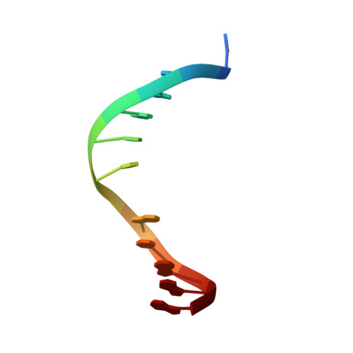
Structure and Specificity of the Vertebrate Anti-Mutator Uracil-DNA Glycosylase Smug1

Wibley, J.E.A.Waters, T.R.Haushalter, K.Verdine, G.L.Pearl, L.H.

(2003) Mol Cell 11: 1647

  • DOI: https://doi.org/10.1016/s1097-2765(03)00235-1
  • Primary Citation of Related Structures:  
    1OE4, 1OE5, 1OE6

  • PubMed Abstract: 

    Cytosine deamination is a major promutagenic process, generating G:U mismatches that can cause transition mutations if not repaired. Uracil is also introduced into DNA via nonmutagenic incorporation of dUTP during replication. In bacteria, uracil is excised by uracil-DNA glycosylases (UDG) related to E. coli UNG, and UNG homologs are found in mammals and viruses. Ung knockout mice display no increase in mutation frequency due to a second UDG activity, SMUG1, which is specialized for antimutational uracil excision in mammalian cells. Remarkably, SMUG1 also excises the oxidation-damage product 5-hydroxymethyluracil (HmU), but like UNG is inactive against thymine (5-methyluracil), a chemical substructure of HmU. We have solved the crystal structure of SMUG1 complexed with DNA and base-excision products. This structure indicates a more invasive interaction with dsDNA than observed with other UDGs and reveals an elegant water displacement/replacement mechanism that allows SMUG1 to exclude thymine from its active site while accepting HmU.


  • Organizational Affiliation

    Cancer Research UK DNA Repair Enzyme Group, Section of Structural Biology, The Institute of Cancer Research, Chester Beatty Laboratories, 237 Fulham Road, London SW3 6JB, United Kingdom.


Macromolecules

Find similar proteins by:  (by identity cutoff)  |  3D Structure
Entity ID: 1
MoleculeChains Sequence LengthOrganismDetailsImage
SINGLE-STRAND SELECTIVE MONOFUNCTIONAL URACIL DNA GLYCOSYLASE
A, B
247Xenopus laevisMutation(s): 0 
EC: 3.2.2
UniProt
Find proteins for Q9YGN6 (Xenopus laevis)
Explore Q9YGN6 
Go to UniProtKB:  Q9YGN6
Entity Groups  
Sequence Clusters30% Identity50% Identity70% Identity90% Identity95% Identity100% Identity
UniProt GroupQ9YGN6
Sequence Annotations
Expand
  • Reference Sequence

Find similar nucleic acids by:  Sequence   |   3D Structure  

Entity ID: 2
MoleculeChains LengthOrganismImage
5'-D(*CP*CP*CP*GP*TP*GP*AP*GP*TP*CP*CP*G)-3'C [auth E]12N/A
Sequence Annotations
Expand
  • Reference Sequence

Find similar nucleic acids by:  Sequence   |   3D Structure  

Entity ID: 3
MoleculeChains LengthOrganismImage
5'-D(*CP*GP*GP*AP*CP*TP*3DR*AP*CP*GP*GP*G)-3'D [auth F]12N/A
Sequence Annotations
Expand
  • Reference Sequence
Experimental Data & Validation

Experimental Data

  • Method: X-RAY DIFFRACTION
  • Resolution: 2.00 Å
  • R-Value Free: 0.216 
  • R-Value Work: 0.177 
  • R-Value Observed: 0.179 
  • Space Group: C 1 2 1
Unit Cell:
Length ( Å )Angle ( ˚ )
a = 121.681α = 90
b = 85.936β = 118.36
c = 79.126γ = 90
Software Package:
Software NamePurpose
REFMACrefinement
MOSFLMdata reduction
SCALAdata scaling
SOLVE/RESOLVEphasing

Structure Validation

View Full Validation Report


Entry History 

Deposition Data

Revision History  (Full details and data files)

  • Version 1.0: 2003-07-11
    Type: Initial release
  • Version 1.1: 2013-07-17
    Changes: Atomic model, Database references, Derived calculations, Non-polymer description, Other, Structure summary, Version format compliance