1OBF

The crystal structure of Glyceraldehyde 3-phosphate Dehydrogenase from Alcaligenes xylosoxidans at 1.7A resolution.


Experimental Data Snapshot

  • Method: X-RAY DIFFRACTION
  • Resolution: 1.70 Å
  • R-Value Free: 0.187 
  • R-Value Work: 0.166 
  • R-Value Observed: 0.167 

wwPDB Validation   3D Report Full Report


This is version 1.2 of the entry. See complete history


Literature

The Structure of Glyceraldehyde 3-Phosphate Dehydrogenase from Alcaligenes Xylosoxidans at 1.7 A Resolution

Antonyuk, S.V.Eady, R.R.Strange, R.W.Hasnain, S.S.

(2003) Acta Crystallogr D Biol Crystallogr 59: 835

  • DOI: https://doi.org/10.1107/s0907444903041441
  • Primary Citation of Related Structures:  
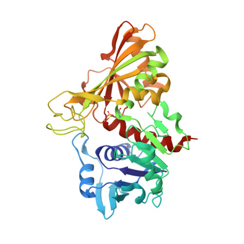
    1OBF

  • PubMed Abstract: 

    The enzyme glyceraldehyde-3-phosphate dehydrogenase (GAPDH) from the Gram-negative denitrifying bacterial species Alcaligenes xylosoxidans was purified and crystallized as a contaminant protein during purification of nitrous oxide reductase. This is the first structure of a GAPDH from a denitrifying species. The crystal structure was solved at 1.7 A resolution by molecular replacement using the structure of GAPDH from Bacillus stearothermophilus as a starting model. The quality of the structure enabled the amino-acid sequence of the A. xylosoxidans GAPDH to be assigned. The structure is that of the apo-enzyme, lacking the NAD+ cofactor and with the active-site residue Cys154 oxidized. The global structure of the enzyme has a homotetrameric quaternary structure similar to that observed for its bacterial and eukaryotic counterparts. The essential role of Cys154 in the enzyme activity has been confirmed. In monomer O two half-occupancy sulfate ions were found at the active site, which are analogous to the substrate and the "attacking" phosphate seen in B. stearothermophilus. One half-occupancy sulfate ion is also located in the substrate-binding site of monomer P.


  • Organizational Affiliation

    Molecular Biophysics Group, CCLRC Daresbury Laboratory, Warrington, Cheshire WA4 4AD, England.


Macromolecules
Find similar proteins by:  (by identity cutoff)  |  3D Structure
Entity ID: 1
MoleculeChains Sequence LengthOrganismDetailsImage
GLYCERALDEHYDE 3-PHOSPHATE DEHYDROGENASEA [auth O],
B [auth P]
335Achromobacter xylosoxidansMutation(s): 0 
EC: 1.2.1.12
UniProt
Find proteins for P83696 (Alcaligenes xylosoxydans xylosoxydans)
Explore P83696 
Go to UniProtKB:  P83696
Entity Groups  
Sequence Clusters30% Identity50% Identity70% Identity90% Identity95% Identity100% Identity
UniProt GroupP83696
Sequence Annotations
Expand
  • Reference Sequence
Experimental Data & Validation

Experimental Data

  • Method: X-RAY DIFFRACTION
  • Resolution: 1.70 Å
  • R-Value Free: 0.187 
  • R-Value Work: 0.166 
  • R-Value Observed: 0.167 
  • Space Group: C 2 2 21
Unit Cell:
Length ( Å )Angle ( ˚ )
a = 88.984α = 90
b = 146.622β = 90
c = 146.622γ = 90
Software Package:
Software NamePurpose
REFMACrefinement
DENZOdata reduction
SCALEPACKdata scaling
MOLREPphasing

Structure Validation

View Full Validation Report


Entry History 

Deposition Data

Revision History  (Full details and data files)

  • Version 1.0: 2003-06-12
    Type: Initial release
  • Version 1.1: 2014-11-05
    Changes: Atomic model, Derived calculations, Non-polymer description, Other, Structure summary, Version format compliance
  • Version 1.2: 2023-12-13
    Changes: Data collection, Database references, Derived calculations, Other, Refinement description