1OAP

Mad structure of the periplasmique domain of the Escherichia coli PAL protein


Experimental Data Snapshot

  • Method: X-RAY DIFFRACTION
  • Resolution: 1.93 Å
  • R-Value Free: 0.234 
  • R-Value Work: 0.200 
  • R-Value Observed: 0.200 

wwPDB Validation   3D Report Full Report


This is version 1.3 of the entry. See complete history


Literature

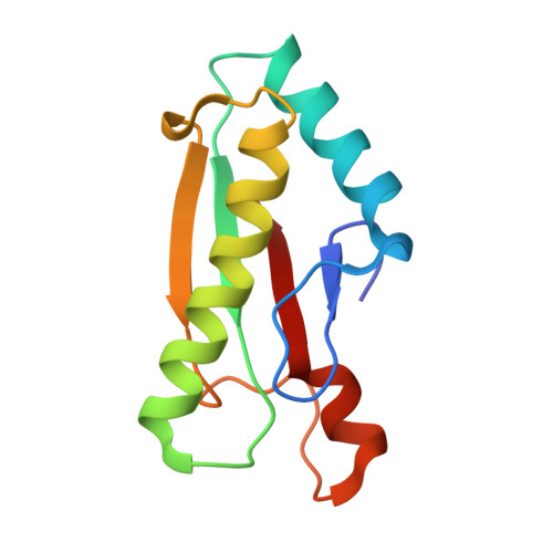
Crystallization and preliminary crystallographic study of the peptidoglycan-associated lipoprotein from Escherichia coli.

Abergel, C.Walburger, A.Chenivesse, S.Lazdunski, C.

(2001) Acta Crystallogr D Biol Crystallogr 57: 317-319

  • DOI: https://doi.org/10.1107/s0907444900019739
  • Primary Citation of Related Structures:  
    1OAP

  • PubMed Abstract: 

    The peptidoglycan-associated lipoprotein (Pal) from Escherichia coli is part of the Tol--Pal multiprotein complex used by group A colicins to penetrate and kill cells. Pal homologues are found in many Gram-negative bacteria and the Tol--Pal system is thought to play a role in bacterial envelope integrity. The Pal protein comprises 152 amino acids. Crystals of the C-terminal 109-amino-acid fragment of the Pal protein have been produced. The crystals belong to the tetragonal space group I4(1), with unit-cell parameters a = b = 89.3, c = 67.2 A. There are two molecules in the asymmetric unit. Frozen crystals diffract to at least 2.8 A resolution using synchrotron radiation. Selenomethionine-substituted truncated Pal protein is currently being produced in order to use multiwavelength anomalous dispersion (MAD) for phasing.


  • Organizational Affiliation

    Information Génétique et Structurale, UMR1889 CNRS-AVENTIS, 31 Chemin Joseph Aiguier, 13402 Marseille CEDEX 20, France. chantal@igs.cnrs-mrs.fr


Macromolecules
Find similar proteins by:  (by identity cutoff)  |  3D Structure
Entity ID: 1
MoleculeChains Sequence LengthOrganismDetailsImage
PEPTIDOGLYCAN-ASSOCIATED LIPOPROTEIN109Escherichia coliMutation(s): 0 
UniProt
Find proteins for P0A912 (Escherichia coli (strain K12))
Explore P0A912 
Go to UniProtKB:  P0A912
Entity Groups  
Sequence Clusters30% Identity50% Identity70% Identity90% Identity95% Identity100% Identity
UniProt GroupP0A912
Sequence Annotations
Expand
  • Reference Sequence
Experimental Data & Validation

Experimental Data

  • Method: X-RAY DIFFRACTION
  • Resolution: 1.93 Å
  • R-Value Free: 0.234 
  • R-Value Work: 0.200 
  • R-Value Observed: 0.200 
  • Space Group: I 41 2 2
Unit Cell:
Length ( Å )Angle ( ˚ )
a = 88.575α = 90
b = 88.575β = 90
c = 68.028γ = 90
Software Package:
Software NamePurpose
MOSFLMdata reduction
SCALAdata scaling
SOLVEphasing
SHARPphasing
CNSrefinement

Structure Validation

View Full Validation Report


Entry History 

Deposition Data

Revision History  (Full details and data files)

  • Version 1.0: 2004-02-13
    Type: Initial release
  • Version 1.1: 2014-02-05
    Changes: Database references, Derived calculations, Non-polymer description, Other, Refinement description, Version format compliance
  • Version 1.2: 2018-03-28
    Changes: Source and taxonomy
  • Version 1.3: 2019-10-09
    Changes: Data collection, Database references, Other