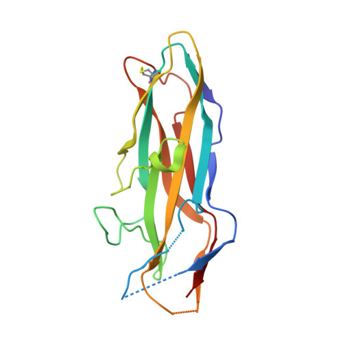
1O9V

F17-aG lectin domain from Escherichia coli in complex with a selenium carbohydrate derivative


Experimental Data Snapshot

  • Method: X-RAY DIFFRACTION
  • Resolution: 1.75 Å
  • R-Value Free: 0.235 
  • R-Value Work: 0.205 
  • R-Value Observed: 0.205 

wwPDB Validation   3D Report Full Report


Ligand Structure Quality Assessment 


This is version 1.4 of the entry. See complete history


Literature

The Fimbrial Adhesin F17-G of Enterotoxigenic Escherichia Coli Has an Immunoglobulin-Like Lectin Domain that Binds N-Acetylglucosamine

Buts, L.Bouckaert, J.De Genst, E.Loris, R.Oscarson, S.Lahmann, M.Messens, J.Brosens, E.Wyns, L.De Greve, H.

(2003) Mol Microbiol 49: 705

  • DOI: https://doi.org/10.1046/j.1365-2958.2003.03600.x
  • Primary Citation of Related Structures:  
    1O9V, 1O9Z

  • PubMed Abstract: 

    The F17-G adhesin at the tip of flexible F17 fimbriae of enterotoxigenic Escherichia coli mediates binding to N-acetyl-beta-D-glucosamine-presenting receptors on the microvilli of the intestinal epithelium of ruminants. We report the 1.7 A resolution crystal structure of the lectin domain of F17-G, both free and in complex with N-acetylglucosamine. The monosaccharide is bound on the side of the ellipsoid-shaped protein in a conserved site around which all natural variations of F17-G are clustered. A model is proposed for the interaction between F17-fimbriated E. coli and microvilli with enhanced affinity compared with the binding constant we determined for F17-G binding to N-acetylglucosamine (0.85 mM-1). Unexpectedly, the F17-G structure reveals that the lectin domains of the F17-G, PapGII and FimH fimbrial adhesins all share the immunoglobulin-like fold of the structural components (pilins) of their fimbriae, despite lack of any sequence identity. Fold comparisons with pilin and chaperone structures of the chaperone/usher pathway highlight the central role of the C-terminal beta-strand G of the immunoglobulin-like fold and provides new insights into pilus assembly, function and adhesion.


  • Organizational Affiliation

    Department of Ultrastructure, Institute for Molecular Biology, Vrije Universiteit Brussel, Vlaams Interuniversitair Instituut voor Biotechnologie, Brussels, Belgium.


Macromolecules
Find similar proteins by:  (by identity cutoff)  |  3D Structure
Entity ID: 1
MoleculeChains Sequence LengthOrganismDetailsImage
F17-AG LECTIN DOMAIN177Escherichia coliMutation(s): 0 
UniProt
Find proteins for Q99003 (Escherichia coli)
Explore Q99003 
Go to UniProtKB:  Q99003
Entity Groups  
Sequence Clusters30% Identity50% Identity70% Identity90% Identity95% Identity100% Identity
UniProt GroupQ99003
Sequence Annotations
Expand
  • Reference Sequence
Small Molecules
Ligands 1 Unique
IDChains Name / Formula / InChI Key2D Diagram3D Interactions
SNG
Query on SNG

Download Ideal Coordinates CCD File 
B [auth A]methyl 2-acetamido-2-deoxy-1-seleno-beta-D-glucopyranoside
C9 H17 N O5 Se
AZZZNYGPGINRNT-OKNNCHMLSA-N
Experimental Data & Validation

Experimental Data

  • Method: X-RAY DIFFRACTION
  • Resolution: 1.75 Å
  • R-Value Free: 0.235 
  • R-Value Work: 0.205 
  • R-Value Observed: 0.205 
  • Space Group: P 61 2 2
Unit Cell:
Length ( Å )Angle ( ˚ )
a = 42.757α = 90
b = 42.757β = 90
c = 273.758γ = 120
Software Package:
Software NamePurpose
CNSrefinement
DENZOdata reduction
SCALEPACKdata scaling
SOLVEphasing

Structure Validation

View Full Validation Report


Ligand Structure Quality Assessment 


Entry History 

Deposition Data

Revision History  (Full details and data files)

  • Version 1.0: 2003-05-29
    Type: Initial release
  • Version 1.1: 2011-05-08
    Changes: Version format compliance
  • Version 1.2: 2011-07-13
    Changes: Version format compliance
  • Version 1.3: 2019-07-24
    Changes: Data collection
  • Version 1.4: 2020-07-29
    Type: Remediation
    Reason: Carbohydrate remediation
    Changes: Derived calculations, Other, Structure summary