1O94

Ternary complex between trimethylamine dehydrogenase and electron transferring flavoprotein


Experimental Data Snapshot

  • Method: X-RAY DIFFRACTION
  • Resolution: 2.00 Å
  • R-Value Free: 0.212 
  • R-Value Work: 0.172 
  • R-Value Observed: 0.174 

wwPDB Validation   3D Report Full Report


Ligand Structure Quality Assessment 


This is version 1.2 of the entry. See complete history


Literature

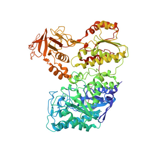
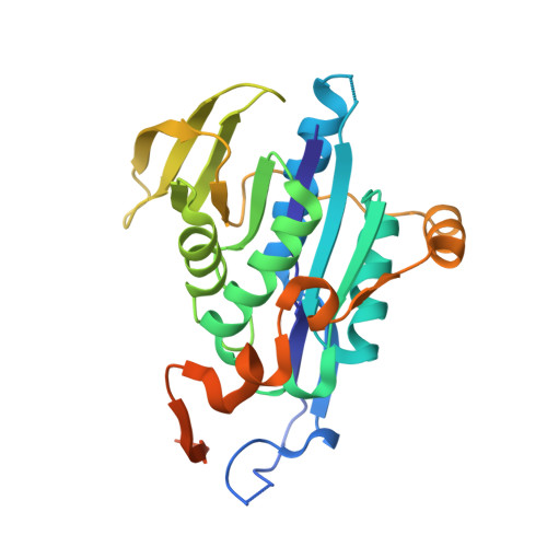
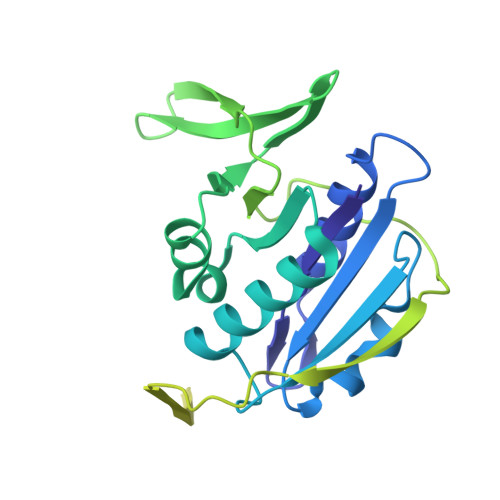
Extensive Conformational Sampling in a Ternary Electron Transfer Complex.

Leys, D.Basran, J.Talfournier, F.Sutcliffe, M.J.Scrutton, N.S.

(2003) Nat Struct Biol 10: 219

  • DOI: https://doi.org/10.1038/nsb894
  • Primary Citation of Related Structures:  
    1O94, 1O95, 1O96, 1O97

  • PubMed Abstract: 

    Here we report the crystal structures of a ternary electron transfer complex showing extensive motion at the protein interface. This physiological complex comprises the iron-sulfur flavoprotein trimethylamine dehydrogenase and electron transferring flavoprotein (ETF) from Methylophilus methylotrophus. In addition, we report the crystal structure of free ETF. In the complex, electron density for the FAD domain of ETF is absent, indicating high mobility. Positions for the FAD domain are revealed by molecular dynamics simulation, consistent with crystal structures and kinetic data. A dual interaction of ETF with trimethylamine dehydrogenase provides for dynamical motion at the protein interface: one site acts as an anchor, thereby allowing the other site to sample a large range of interactions, some compatible with rapid electron transfer. This study establishes the role of conformational sampling in multi-domain redox systems, providing insight into electron transfer between ETFs and structurally distinct redox partners.


  • Organizational Affiliation

    Department of Biochemistry, University of Leicester, University Road, Leicester LE1 7RH, UK. dl37@le.ac.uk


Macromolecules
Find similar proteins by:  (by identity cutoff)  |  3D Structure
Entity ID: 1
MoleculeChains Sequence LengthOrganismDetailsImage
TRIMETHYLAMINE DEHYDROGENASE
A, B
729Methylophilus methylotrophusMutation(s): 0 
EC: 1.5.99.7
UniProt
Find proteins for P16099 (Methylophilus methylotrophus)
Explore P16099 
Go to UniProtKB:  P16099
Entity Groups  
Sequence Clusters30% Identity50% Identity70% Identity90% Identity95% Identity100% Identity
UniProt GroupP16099
Sequence Annotations
Expand
  • Reference Sequence
Find similar proteins by:  (by identity cutoff)  |  3D Structure
Entity ID: 2
MoleculeChains Sequence LengthOrganismDetailsImage
ELECTRON TRANSFER FLAVOPROTEIN BETA-SUBUNIT
C, E
264Methylophilus methylotrophusMutation(s): 0 
UniProt
Find proteins for P53570 (Methylophilus methylotrophus)
Explore P53570 
Go to UniProtKB:  P53570
Entity Groups  
Sequence Clusters30% Identity50% Identity70% Identity90% Identity95% Identity100% Identity
UniProt GroupP53570
Sequence Annotations
Expand
  • Reference Sequence
Find similar proteins by:  (by identity cutoff)  |  3D Structure
Entity ID: 3
MoleculeChains Sequence LengthOrganismDetailsImage
ELECTRON TRANSFER FLAVOPROTEIN ALPHA-SUBUNIT
D, F
320Methylophilus methylotrophusMutation(s): 0 
UniProt
Find proteins for P53571 (Methylophilus methylotrophus)
Explore P53571 
Go to UniProtKB:  P53571
Entity Groups  
Sequence Clusters30% Identity50% Identity70% Identity90% Identity95% Identity100% Identity
UniProt GroupP53571
Sequence Annotations
Expand
  • Reference Sequence
Small Molecules
Ligands 4 Unique
IDChains Name / Formula / InChI Key2D Diagram3D Interactions
FMN
Query on FMN

Download Ideal Coordinates CCD File 
G [auth A],
J [auth B]
FLAVIN MONONUCLEOTIDE
C17 H21 N4 O9 P
FVTCRASFADXXNN-SCRDCRAPSA-N
ADP
Query on ADP

Download Ideal Coordinates CCD File 
H [auth A],
K [auth B]
ADENOSINE-5'-DIPHOSPHATE
C10 H15 N5 O10 P2
XTWYTFMLZFPYCI-KQYNXXCUSA-N
SF4
Query on SF4

Download Ideal Coordinates CCD File 
I [auth A],
L [auth B]
IRON/SULFUR CLUSTER
Fe4 S4
LJBDFODJNLIPKO-UHFFFAOYSA-N
AMP
Query on AMP

Download Ideal Coordinates CCD File 
M [auth C],
N [auth E]
ADENOSINE MONOPHOSPHATE
C10 H14 N5 O7 P
UDMBCSSLTHHNCD-KQYNXXCUSA-N
Experimental Data & Validation

Experimental Data

  • Method: X-RAY DIFFRACTION
  • Resolution: 2.00 Å
  • R-Value Free: 0.212 
  • R-Value Work: 0.172 
  • R-Value Observed: 0.174 
  • Space Group: C 1 2 1
Unit Cell:
Length ( Å )Angle ( ˚ )
a = 118.93α = 90
b = 211.543β = 100.03
c = 125.97γ = 90
Software Package:
Software NamePurpose
REFMACrefinement
DENZOdata reduction
SCALEPACKdata scaling
AMoREphasing

Structure Validation

View Full Validation Report


Ligand Structure Quality Assessment 


Entry History 

Deposition Data

Revision History  (Full details and data files)

  • Version 1.0: 2003-02-06
    Type: Initial release
  • Version 1.1: 2013-09-18
    Changes: Derived calculations, Non-polymer description, Other, Version format compliance
  • Version 1.2: 2023-12-13
    Changes: Data collection, Database references, Derived calculations, Other, Refinement description