1O5W

The structure basis of specific recognitions for substrates and inhibitors of rat monoamine oxidase A


Experimental Data Snapshot

  • Method: X-RAY DIFFRACTION
  • Resolution: 3.20 Å
  • R-Value Free: 0.282 
  • R-Value Work: 0.220 
  • R-Value Observed: 0.242 

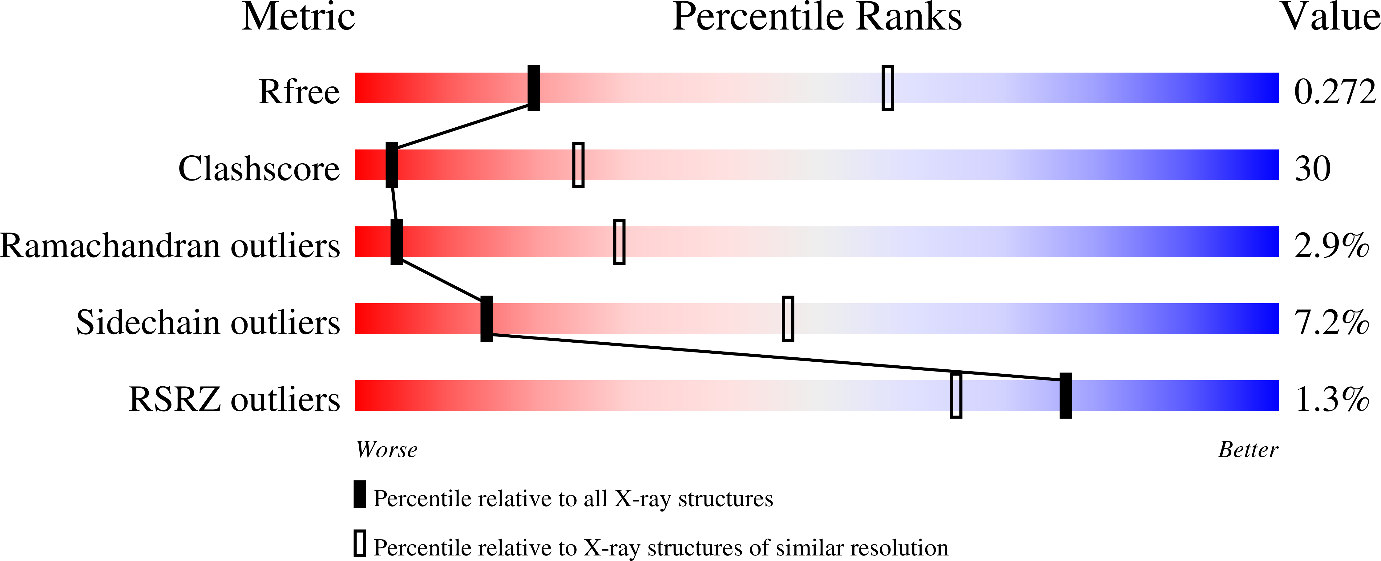
wwPDB Validation   3D Report Full Report


Ligand Structure Quality Assessment 


This is version 1.3 of the entry. See complete history


Literature

Structure of rat monoamine oxidase a and its specific recognitions for substrates and inhibitors.

Ma, J.Yoshimura, M.Yamashita, E.Nakagawa, A.Ito, A.Tsukihara, T.

(2004) J Mol Biol 338: 103-114

  • DOI: https://doi.org/10.1016/j.jmb.2004.02.032
  • Primary Citation of Related Structures:  
    1O5W

  • PubMed Abstract: 

    Monoamine oxidase (MAO), a mitochondrial outer membrane enzyme, catalyzes the degradation of neurotransmitters in the central nervous system and is the target for anti-depression drug design. Two subtypes of MAO, MAOA and MAOB, are similar in primary sequences but have unique substrate and inhibitor specificities. The structures of human MAOB complexed with various inhibitors were reported early. To understand the mechanisms of specific substrate and inhibitor recognitions of MAOA and MAOB, we have determined the crystal structure of rat MAOA complexed with the specific inhibitor, clorgyline, at 3.2A resolution. The comparison of the structures between MAOA and MAOB clearly explains the specificity of clorgyline for MAOA inhibition. The fitting of serotonin into the binding pockets of MAOs demonstrates that MAOB Tyr326 would block access of the 5-hydroxy group of serotonin into the enzyme. These results will lead to further understanding of the MAOA function and to new anti-depression drug design. This study also presents that MAOA has a transmembrane helix at the C-terminal region. This is the first crystal structure of membrane protein with an isolated transmembrane helix.


  • Organizational Affiliation

    Institute for Protein Research, Osaka University, Yamadaoka 3-2, Suita, Osaka 565-0871, Japan.


Macromolecules
Find similar proteins by:  (by identity cutoff)  |  3D Structure
Entity ID: 1
MoleculeChains Sequence LengthOrganismDetailsImage
Amine oxidase [flavin-containing] A
A, B, C, D
534Rattus norvegicusMutation(s): 0 
EC: 1.4.3.4
Membrane Entity: Yes 
UniProt
Find proteins for P21396 (Rattus norvegicus)
Explore P21396 
Go to UniProtKB:  P21396
Entity Groups  
Sequence Clusters30% Identity50% Identity70% Identity90% Identity95% Identity100% Identity
UniProt GroupP21396
Sequence Annotations
Expand
  • Reference Sequence
Small Molecules
Binding Affinity Annotations 
IDSourceBinding Affinity
MLG BindingDB:  1O5W Ki: min: 12, max: 1200 (nM) from 5 assay(s)
IC50: min: 4.90e-3, max: 48 (nM) from 52 assay(s)
Experimental Data & Validation

Experimental Data

  • Method: X-RAY DIFFRACTION
  • Resolution: 3.20 Å
  • R-Value Free: 0.282 
  • R-Value Work: 0.220 
  • R-Value Observed: 0.242 
  • Space Group: P 43 21 2
Unit Cell:
Length ( Å )Angle ( ˚ )
a = 157.56α = 90
b = 157.56β = 90
c = 257.84γ = 90
Software Package:
Software NamePurpose
CNSrefinement
MOSFLMdata reduction
CCP4data scaling
AMoREphasing

Structure Validation

View Full Validation Report


Ligand Structure Quality Assessment 


Entry History 

Deposition Data

Revision History  (Full details and data files)

  • Version 1.0: 2004-04-20
    Type: Initial release
  • Version 1.1: 2008-04-27
    Changes: Version format compliance
  • Version 1.2: 2011-07-13
    Changes: Version format compliance
  • Version 1.3: 2023-12-27
    Changes: Data collection, Database references, Derived calculations