1O1S

Structure of FPT bound to isoprenoid analog 3b


Experimental Data Snapshot

  • Method: X-RAY DIFFRACTION
  • Resolution: 2.30 Å
  • R-Value Work: 0.190 
  • R-Value Observed: 0.190 

wwPDB Validation   3D Report Full Report


This is version 1.5 of the entry. See complete history


Literature

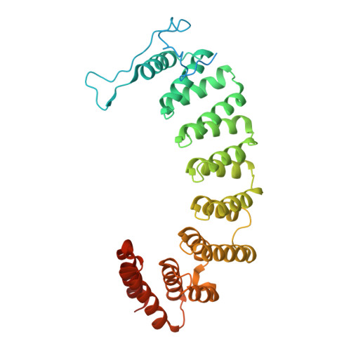
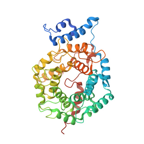
Biochemical and Structural Studies with Prenyl Diphosphate Analogues Provide Insights into Isoprenoid Recognition by Protein Farnesyl Transferase

Turek-Etienne, T.C.Strickland, C.L.Distefano, M.D.

(2003) Biochemistry 42: 3716-3724

  • DOI: https://doi.org/10.1021/bi0266838
  • Primary Citation of Related Structures:  
    1O1R, 1O1S, 1O1T

  • PubMed Abstract: 

    Protein farnesyl transferase (PFTase) catalyzes the reaction between farnesyl diphosphate and a protein substrate to form a thioether-linked prenylated protein. The fact that many prenylated proteins are involved in signaling processes has generated considerable interest in protein prenyl transferases as possible anticancer targets. While considerable progress has been made in understanding how prenyl transferases distinguish between related target proteins, the rules for isoprenoid discrimination by these enzymes are less well understood. To clarify how PFTase discriminates between FPP and larger prenyl diphosphates, we have examined the interactions between the enzyme and several isoprenoid analogues, GGPP, and the farnesylated peptide product using a combination of biochemical and structural methods. Two photoactive isoprenoid analogues were shown to inhibit yeast PFTase with K(I) values as low as 45 nM. Crystallographic analysis of one of these analogues bound to PFTase reveals that the diphosphate moiety and the two isoprene units bind in the same positions occupied by the corresponding atoms in FPP when bound to PFTase. However, the benzophenone group protrudes into the acceptor protein binding site and prevents the binding of the second (protein) substrate. Crystallographic analysis of geranylgeranyl diphosphate bound to PFTase shows that the terminal two isoprene units and diphosphate group of the molecule map to the corresponding atoms in FPP; however, the first and second isoprene units bulge away from the acceptor protein binding site. Comparison of the GGPP binding mode with the binding of the farnesylated peptide product suggests that the bulkier isoprenoid cannot rearrange to convert to product without unfavorable steric interactions with the acceptor protein. Taken together, these data do not support the "molecular ruler hypotheses". Instead, we propose a "second site exclusion model" in which PFTase binds larger isoprenoids in a fashion that prevents the subsequent productive binding of the acceptor protein or its conversion to product.


  • Organizational Affiliation

    Department of Chemistry, University of Minnesota, Minneapolis, Minnesota 55455, USA.


Macromolecules
Find similar proteins by:  (by identity cutoff)  |  3D Structure
Entity ID: 1
MoleculeChains Sequence LengthOrganismDetailsImage
Protein farnesyltransferase alpha subunit380Rattus norvegicusMutation(s): 0 
EC: 2.5.1
UniProt
Find proteins for Q04631 (Rattus norvegicus)
Explore Q04631 
Go to UniProtKB:  Q04631
Entity Groups  
Sequence Clusters30% Identity50% Identity70% Identity90% Identity95% Identity100% Identity
UniProt GroupQ04631
Sequence Annotations
Expand
  • Reference Sequence
Find similar proteins by:  (by identity cutoff)  |  3D Structure
Entity ID: 2
MoleculeChains Sequence LengthOrganismDetailsImage
Protein farnesyltransferase beta subunit427Rattus norvegicusMutation(s): 0 
EC: 2.5.1
UniProt
Find proteins for Q02293 (Rattus norvegicus)
Explore Q02293 
Go to UniProtKB:  Q02293
Entity Groups  
Sequence Clusters30% Identity50% Identity70% Identity90% Identity95% Identity100% Identity
UniProt GroupQ02293
Sequence Annotations
Expand
  • Reference Sequence
Small Molecules
Ligands 2 Unique
IDChains Name / Formula / InChI Key2D Diagram3D Interactions
1NH
Query on 1NH

Download Ideal Coordinates CCD File 
D [auth B](2E,6E)-8-[(3-BENZOYLBENZYL)OXY]-3,7-DIMETHYLOCTA-2,6-DIENYL TRIHYDROGEN DIPHOSPHATE
C24 H30 O9 P2
IRMXEEMZDGNJKM-APFDVHGWSA-N
ZN
Query on ZN

Download Ideal Coordinates CCD File 
C [auth B]ZINC ION
Zn
PTFCDOFLOPIGGS-UHFFFAOYSA-N
Binding Affinity Annotations 
IDSourceBinding Affinity
1NH Binding MOAD:  1O1S Ki: 49 (nM) from 1 assay(s)
PDBBind:  1O1S Ki: 49 (nM) from 1 assay(s)
Experimental Data & Validation

Experimental Data

  • Method: X-RAY DIFFRACTION
  • Resolution: 2.30 Å
  • R-Value Work: 0.190 
  • R-Value Observed: 0.190 
  • Space Group: P 61
Unit Cell:
Length ( Å )Angle ( ˚ )
a = 171.83α = 90
b = 171.83β = 90
c = 69.09γ = 120
Software Package:
Software NamePurpose
DENZOdata reduction
X-PLORmodel building
X-PLORrefinement
SCALEPACKdata scaling
X-PLORphasing

Structure Validation

View Full Validation Report


Entry History 

Deposition Data

Revision History  (Full details and data files)

  • Version 1.0: 2003-05-06
    Type: Initial release
  • Version 1.1: 2008-04-26
    Changes: Version format compliance
  • Version 1.2: 2011-07-13
    Changes: Version format compliance
  • Version 1.3: 2017-10-04
    Changes: Advisory, Refinement description
  • Version 1.4: 2023-12-27
    Changes: Advisory, Data collection, Database references, Derived calculations
  • Version 1.5: 2024-03-13
    Changes: Source and taxonomy, Structure summary