1O07

Crystal Structure of the complex between Q120L/Y150E mutant of AmpC and a beta-lactam inhibitor (MXG)


Experimental Data Snapshot

  • Method: X-RAY DIFFRACTION
  • Resolution: 1.71 Å
  • R-Value Free: 0.191 
  • R-Value Work: 0.154 
  • R-Value Observed: 0.156 

wwPDB Validation   3D Report Full Report


Ligand Structure Quality Assessment 


This is version 1.5 of the entry. See complete history


Literature

Structural Aspects for Evolution of beta-Lactamases from Penicillin-Binding Proteins

Meroueh, S.O.Minasov, G.Lee, W.Shoichet, B.K.Mobashery, S.

(2003) J Am Chem Soc 125: 9612-9618

  • DOI: https://doi.org/10.1021/ja034861u
  • Primary Citation of Related Structures:  
    1O07

  • PubMed Abstract: 

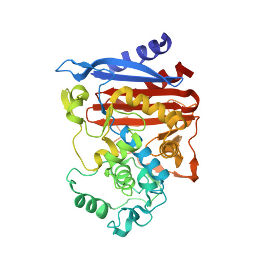
    Penicillin-binding proteins (PBPs), biosynthetic enzymes of bacterial cell wall assembly, and beta-lactamases, resistance enzymes to beta-lactam antibiotics, are related to each other from an evolutionary point of view. Massova and Mobashery (Antimicrob. Agents Chemother. 1998, 42, 1-17) have proposed that for beta-lactamases to have become effective at their function as antibiotic resistance enzymes, they would have had to undergo structure alterations such that they would not interact with the peptidoglycan, which is the substrate for PBPs. A cephalosporin analogue, 7beta-[N-Acetyl-L-alanyl-gamma-D-glutamyl-L-lysine]-3-acetoxymethyl-3-cephem-carboxylic acid (compound 6), was conceived and synthesized to test this notion. The X-ray structure of the complex of this cephalosporin bound to the active site of the deacylation-deficient Q120L/Y150E variant of the class C AmpC beta-lactamase from Escherichia coli was solved at 1.71 A resolution. This complex revealed that the surface for interaction with the strand of peptidoglycan that acylates the active site, which is present in PBPs, is absent in the -lactamase active site. Furthermore, insertion of a peptide in the beta-lactamase active site at a location where the second strand of peptidoglycan in some PBPs binds has effectively abolished the possibility for such interaction with the beta-lactamase. A 2.6 ns dynamics simulation was carried out for the complex, which revealed that the peptidoglycan surrogate (i.e., the active-site-bound ligand) undergoes substantial motion and is not stabilized for binding within the active site. These factors taken together disclose the set of structure modifications in the antibiotic resistance enzyme that prevent it from interacting with the peptidoglycan, en route to achieving catalytic proficiency for their intended function.


  • Organizational Affiliation

    Department of Chemistry and Biochemistry, University of Notre Dame, Notre Dame, Indiana 46556, USA.


Macromolecules
Find similar proteins by:  (by identity cutoff)  |  3D Structure
Entity ID: 1
MoleculeChains Sequence LengthOrganismDetailsImage
Beta-lactamase
A, B
358Escherichia coliMutation(s): 2 
EC: 3.5.2.6
UniProt
Find proteins for P00811 (Escherichia coli (strain K12))
Explore P00811 
Go to UniProtKB:  P00811
Entity Groups  
Sequence Clusters30% Identity50% Identity70% Identity90% Identity95% Identity100% Identity
UniProt GroupP00811
Sequence Annotations
Expand
  • Reference Sequence
Small Molecules
Ligands 2 Unique
IDChains Name / Formula / InChI Key2D Diagram3D Interactions
MXG
Query on MXG

Download Ideal Coordinates CCD File 
D [auth A],
F [auth B]
2-(1-{2-[4-(2-ACETYLAMINO-PROPIONYLAMINO)-4-CARBOXY-BUTYRYLAMINO]-6-AMINO-HEXANOYLAMINO}-2-OXO-ETHYL)-5-METHYLENE-5,6-DIHYDRO-2H-[1,3]THIAZINE-4-CARBOXYLIC ACID
C24 H36 N6 O9 S
BZBLEHRBZDVRSG-JKCYHXNASA-N
K
Query on K

Download Ideal Coordinates CCD File 
C [auth A],
E [auth B]
POTASSIUM ION
K
NPYPAHLBTDXSSS-UHFFFAOYSA-N
Experimental Data & Validation

Experimental Data

  • Method: X-RAY DIFFRACTION
  • Resolution: 1.71 Å
  • R-Value Free: 0.191 
  • R-Value Work: 0.154 
  • R-Value Observed: 0.156 
  • Space Group: C 1 2 1
Unit Cell:
Length ( Å )Angle ( ˚ )
a = 118.808α = 90
b = 76.298β = 116.21
c = 98.127γ = 90
Software Package:
Software NamePurpose
REFMACrefinement
DENZOdata reduction
SCALEPACKdata scaling
AMoREphasing

Structure Validation

View Full Validation Report


Ligand Structure Quality Assessment 


Entry History 

Deposition Data

Revision History  (Full details and data files)

  • Version 1.0: 2003-08-26
    Type: Initial release
  • Version 1.1: 2008-04-29
    Changes: Version format compliance
  • Version 1.2: 2011-07-13
    Changes: Atomic model, Version format compliance
  • Version 1.3: 2021-10-27
    Changes: Database references, Derived calculations
  • Version 1.4: 2024-02-14
    Changes: Data collection
  • Version 1.5: 2024-04-03
    Changes: Refinement description