1NY2

Human alpha thrombin inhibited by RPPGF and hirugen


Experimental Data Snapshot

  • Method: X-RAY DIFFRACTION
  • Resolution: 2.30 Å
  • R-Value Work: 0.202 

wwPDB Validation   3D Report Full Report


This is version 1.6 of the entry. See complete history


Literature

Mechanisms of Arg-Pro-Pro-Gly-Phe inhibition of thrombin.

Hasan, A.A.Warnock, M.Nieman, M.Srikanth, S.Mahdi, F.Krishnan, R.Tulinsky, A.Schmaier, A.H.

(2003) Am J Physiol Heart Circ Physiol 285: H183-H193

  • DOI: https://doi.org/10.1152/ajpheart.00490.2002
  • Primary Citation of Related Structures:  
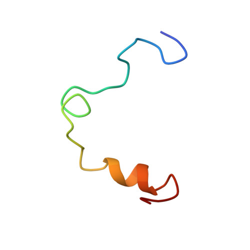
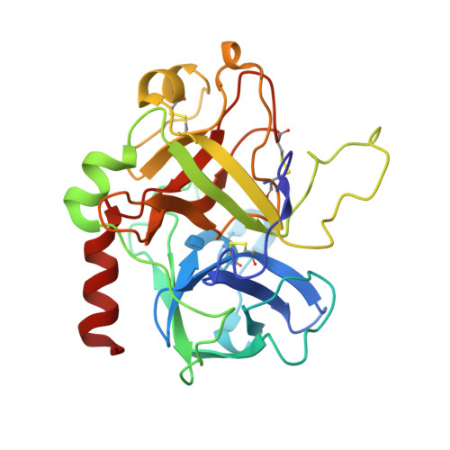
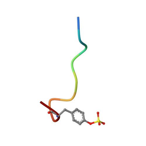
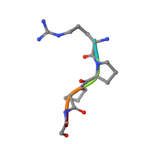
    1NY2

  • PubMed Abstract: 

    Investigations determined the mechanism(s) by which Arg-Pro-Pro-Gly-Phe (RPPGF) inhibits thrombin-induced platelet activation. High concentrations of RPPGF inhibit thrombin-induced coagulant activity. RPPGF binds to the active site of thrombin by forming a parallel beta-strand with Ser214-Gly216 and interacts with His57, Asp189, and Ser195 of the catalytic triad. RPPGF competitively inhibits alpha-thrombin from hydrolyzing Sar-Pro-Arg-paranitroanilide with a Ki = 1.75 +/- 0.03 mM. Other mechanisms were sought to explain why RPPGF inhibits thrombin activation of platelets at concentrations below that which inhibits its active site. Soluble RPPGF blocks biotinylated NATLDPRSFLLR of the thrombin cleavage site on protease-activated receptor (PAR)1 from binding to the peptide RPPGC (IC50 = 20 microM). The soluble recombinant extracellular domain of PAR1 (rPAR1EC) blocks biotinylated RPPGF binding to rPAR1EC (IC50 = 50 microM) bound to microtiter plates, but rPAR1EC deletion mutants missing the sequence LDPR or PRSF do not. RPPGF and related forms prevent the thrombin-like enzyme thrombocytin from proteolyzing rPAR1EC at concentrations that do not block thrombocytin's active site. These studies indicate that RPPGF is a bifunctional inhibitor of thrombin: it binds to PAR1 to prevent thrombin cleavage at Arg41 and interacts with the active site of alpha-thrombin.


  • Organizational Affiliation

    Department of Internal Medicine, University of Michigan, Ann Arbor, MI 48109-0640, USA.


Macromolecules
Find similar proteins by:  (by identity cutoff)  |  3D Structure
Entity ID: 1
MoleculeChains Sequence LengthOrganismDetailsImage
thrombin light chainA [auth 1]36Homo sapiensMutation(s): 0 
EC: 3.4.21.5
UniProt & NIH Common Fund Data Resources
Find proteins for P00734 (Homo sapiens)
Explore P00734 
Go to UniProtKB:  P00734
PHAROS:  P00734
GTEx:  ENSG00000180210 
Entity Groups  
Sequence Clusters30% Identity50% Identity70% Identity90% Identity95% Identity100% Identity
UniProt GroupP00734
Sequence Annotations
Expand
  • Reference Sequence
Find similar proteins by:  (by identity cutoff)  |  3D Structure
Entity ID: 2
MoleculeChains Sequence LengthOrganismDetailsImage
thrombin Heavy chainB [auth 2]259Homo sapiensMutation(s): 0 
EC: 3.4.21.5
UniProt & NIH Common Fund Data Resources
Find proteins for P00734 (Homo sapiens)
Explore P00734 
Go to UniProtKB:  P00734
PHAROS:  P00734
GTEx:  ENSG00000180210 
Entity Groups  
Sequence Clusters30% Identity50% Identity70% Identity90% Identity95% Identity100% Identity
UniProt GroupP00734
Sequence Annotations
Expand
  • Reference Sequence

Find similar proteins by:  Sequence   |   3D Structure  

Entity ID: 3
MoleculeChains Sequence LengthOrganismDetailsImage
HirugenC [auth 3]10N/AMutation(s): 0 
UniProt
Find proteins for P28504 (Hirudo medicinalis)
Explore P28504 
Go to UniProtKB:  P28504
Entity Groups  
Sequence Clusters30% Identity50% Identity70% Identity90% Identity95% Identity100% Identity
UniProt GroupP28504
Sequence Annotations
Expand
  • Reference Sequence

Find similar proteins by:  Sequence   |   3D Structure  

Entity ID: 4
MoleculeChains Sequence LengthOrganismDetailsImage
Inhibitor peptide RPPGFD [auth 4]5N/AMutation(s): 0 
UniProt & NIH Common Fund Data Resources
Find proteins for P01042 (Homo sapiens)
Explore P01042 
Go to UniProtKB:  P01042
PHAROS:  P01042
GTEx:  ENSG00000113889 
Entity Groups  
UniProt GroupP01042
Sequence Annotations
Expand
  • Reference Sequence
Small Molecules
Modified Residues  1 Unique
IDChains TypeFormula2D DiagramParent
TYS
Query on TYS
C [auth 3]L-PEPTIDE LINKINGC9 H11 N O6 STYR
Experimental Data & Validation

Experimental Data

  • Method: X-RAY DIFFRACTION
  • Resolution: 2.30 Å
  • R-Value Work: 0.202 
  • Space Group: P 21 21 2
Unit Cell:
Length ( Å )Angle ( ˚ )
a = 79.15α = 90
b = 104.97β = 90
c = 45.18γ = 90
Software Package:
Software NamePurpose
AMoREphasing
PROLSQrefinement

Structure Validation

View Full Validation Report


Entry History 

Deposition Data

Revision History  (Full details and data files)

  • Version 1.0: 2003-03-04
    Type: Initial release
  • Version 1.1: 2008-04-29
    Changes: Version format compliance
  • Version 1.2: 2011-07-13
    Changes: Atomic model, Database references, Derived calculations, Non-polymer description, Structure summary, Version format compliance
  • Version 1.3: 2015-03-25
    Changes: Database references
  • Version 1.4: 2017-10-11
    Changes: Refinement description
  • Version 1.5: 2023-08-16
    Changes: Data collection, Database references, Derived calculations, Refinement description
  • Version 1.6: 2023-11-15
    Changes: Data collection