1NU2

Crystal structure of the murine Disabled-1 (Dab1) PTB domain-ApoER2 peptide-PI-4,5P2 ternary complex


Experimental Data Snapshot

  • Method: X-RAY DIFFRACTION
  • Resolution: 1.90 Å
  • R-Value Free: 0.253 
  • R-Value Work: 0.204 
  • R-Value Observed: 0.238 

wwPDB Validation   3D Report Full Report


Ligand Structure Quality Assessment 


This is version 1.5 of the entry. See complete history


Literature

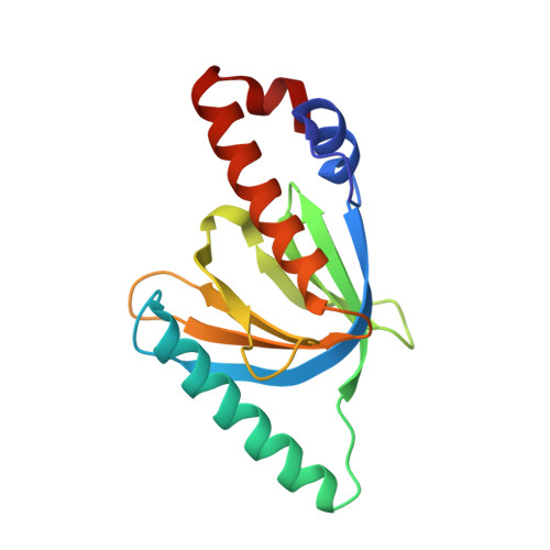
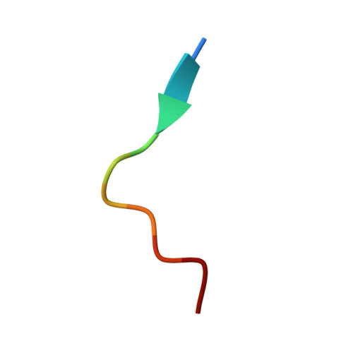
Origins of Peptide Selectivity and Phosphoinositide Binding Revealed by Structures of Disabled-1 PTB Domain Complexes

Stolt, P.C.Jeon, H.Song, H.K.Herz, J.Eck, M.J.Blacklow, S.C.

(2003) Structure 11: 569-579

  • DOI: https://doi.org/10.1016/s0969-2126(03)00068-6
  • Primary Citation of Related Structures:  
    1NTV, 1NU2

  • PubMed Abstract: 

    Formation of the mammalian six-layered neocortex depends on a signaling pathway that involves Reelin, the very low-density lipoprotein receptor, the apolipoprotein E receptor-2 (ApoER2), and the adaptor protein Disabled-1 (Dab1). The 1.5 A crystal structure of a complex between the Dab1 phosphotyrosine binding (PTB) domain and a 14-residue peptide from the ApoER2 tail explains the unusual preference of Dab1 for unphosphorylated tyrosine within the NPxY motif of the peptide. Crystals of the complex soaked with the phosphoinositide PI-4,5P(2) (PI) show that PI binds to conserved basic residues on the PTB domain opposite the peptide binding groove. This finding explains how the Dab1 PTB domain can simultaneously bind PI and the ApoER2 tail. Recruitment of the Dab1 PTB domain to PI-rich regions of the plasma membrane may facilitate association with the Reelin receptor cytoplasmic tails to transduce a critical positional cue to migrating neurons.


  • Organizational Affiliation

    Department of Pathology, Brigham and Women's Hospital and Harvard Medical School, 75 Francis Street, Boston, MA 02115, USA.


Macromolecules
Find similar proteins by:  (by identity cutoff)  |  3D Structure
Entity ID: 1
MoleculeChains Sequence LengthOrganismDetailsImage
Disabled homolog 1152Mus musculusMutation(s): 0 
Gene Names: DAB1
UniProt
Find proteins for P97318 (Mus musculus)
Explore P97318 
Go to UniProtKB:  P97318
Entity Groups  
Sequence Clusters30% Identity50% Identity70% Identity90% Identity95% Identity100% Identity
UniProt GroupP97318
Sequence Annotations
Expand
  • Reference Sequence

Find similar proteins by:  Sequence   |   3D Structure  

Entity ID: 2
MoleculeChains Sequence LengthOrganismDetailsImage
peptide derived from murine Apolipoprotein E Receptor-210N/AMutation(s): 0 
Entity Groups  
Sequence Clusters30% Identity50% Identity70% Identity90% Identity95% Identity100% Identity
Sequence Annotations
Expand
  • Reference Sequence
Small Molecules
Ligands 1 Unique
IDChains Name / Formula / InChI Key2D Diagram3D Interactions
I3P
Query on I3P

Download Ideal Coordinates CCD File 
C [auth A]D-MYO-INOSITOL-1,4,5-TRIPHOSPHATE
C6 H15 O15 P3
MMWCIQZXVOZEGG-XJTPDSDZSA-N
Experimental Data & Validation

Experimental Data

  • Method: X-RAY DIFFRACTION
  • Resolution: 1.90 Å
  • R-Value Free: 0.253 
  • R-Value Work: 0.204 
  • R-Value Observed: 0.238 
  • Space Group: P 21 21 21
Unit Cell:
Length ( Å )Angle ( ˚ )
a = 36.27α = 90
b = 45.984β = 90
c = 89.808γ = 90
Software Package:
Software NamePurpose
DENZOdata reduction
SCALEPACKdata scaling
CNSrefinement
CNSphasing

Structure Validation

View Full Validation Report


Ligand Structure Quality Assessment 


Entry History 

Deposition Data

Revision History  (Full details and data files)

  • Version 1.0: 2003-04-15
    Type: Initial release
  • Version 1.1: 2008-04-29
    Changes: Version format compliance
  • Version 1.2: 2011-07-13
    Changes: Version format compliance
  • Version 1.3: 2018-04-04
    Changes: Data collection
  • Version 1.4: 2023-08-16
    Changes: Data collection, Database references, Derived calculations, Refinement description
  • Version 1.5: 2024-03-13
    Changes: Source and taxonomy, Structure summary