Crystal structure of the sensor domain of the BlaR penicillin receptor from Bacillus licheniformis.
Kerff, F., Charlier, P., Colombo, M.L., Sauvage, E., Brans, A., Frere, J.M., Joris, B., Fonze, E.(2003) Biochemistry 42: 12835-12843
- PubMed: 14596597
- DOI: https://doi.org/10.1021/bi034976a
- Primary Citation of Related Structures:
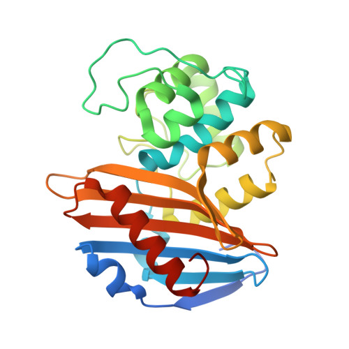
1NRF - PubMed Abstract:
As in several staphylococci, the synthesis of the Bacillus licheniformis 749/I beta-lactamase is an inducible phenomenon regulated by a signal-transducing membrane protein BlaR. The C-terminal domain of this multimodular protein is an extracellular domain which specifically recognizes beta-lactam antibiotics. When it binds a beta-lactam, a signal is transmitted by the transmembrane region to the intracellular loops. In response, the hydrolytic activity of the BlaR large cytoplasmic L3 loop is induced, and a cascade of reactions is generated, leading to the transcription of the beta-lactamase gene. Here, we describe the crystal structure of the extracellular penicillin-receptor domain of BlaR (residues 346-601) at 2.5 A resolution in order to understand why this domain, whose folding is very similar to that of class D beta-lactamases, behaves as a highly sensitive penicillin-binding protein rather than a beta-lactamase. Two residues of the BlaR C-terminal domain, Thr452 and Thr542, modify the hydrophobic characteristic of the class D beta-lactamase active site. Both residues seem to be in part responsible for the lack of beta-lactamase activity of the BlaR protein due to the stability of the acyl-enzyme. Although further experimental data are needed to fully understand the transmembrane induction process, the comparison of the BlaR sensor domain structure with those of class D beta-lactamase complexes and penicillin-binding proteins provides interesting elements to hypothesize on possible signal transmission mechanisms.
Organizational Affiliation:
Institut de Physique B5, Université de Liège, B-4000 Sart Tilman, Belgium.