The 2.3-A crystal structure of the shikimate 5-dehydrogenase orthologue YdiB from Escherichia coli suggests a novel catalytic environment for an NAD-dependent dehydrogenase
Benach, J., Lee, I., Edstrom, W., Kuzin, A.P., Chiang, Y., Acton, T.B., Montelione, G.T., Hunt, J.F.(2003) J Biol Chem 278: 19176-19182
- PubMed: 12624088
- DOI: https://doi.org/10.1074/jbc.M301348200
- Primary Citation of Related Structures:
1NPD - PubMed Abstract:
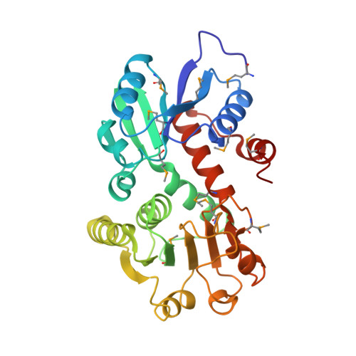
We present here the 2.3-A crystal structure of the Escherichia coli YdiB protein, an orthologue of shikimate 5-dehydrogenase. This enzyme catalyzes the reduction of 3-dehydroshikimate to shikimate as part of the shikimate pathway, which is absent in mammals but required for the de novo synthesis of aromatic amino acids, quinones, and folate in many other organisms. In this context, the shikimate pathway has been promoted as a target for the development of antimicrobial agents. The crystal structure of YdiB shows that the protomer contains two alpha/beta domains connected by two alpha-helices, with the N-terminal domain being novel and the C-terminal domain being a Rossmann fold. The NAD+ cofactor, which co-purified with the enzyme, is bound to the Rossmann domain in an elongated fashion with the nicotinamide ring in the pro-R conformation. Its binding site contains several unusual features, including a cysteine residue in close apposition to the nicotinamide ring and a clamp over the ribose of the adenosine moiety formed by phenylalanine and lysine residues. The structure explains the specificity for NAD versus NADP in different members of the shikimate dehydrogenase family on the basis of variations in the amino acid identity of several other residues in the vicinity of this ribose group. A cavity lined by residues that are 100% conserved among all shikimate dehydrogenases is found between the two domains of YdiB, in close proximity to the hydride acceptor site on the nicotinamide ring. Shikimate was modeled into this site in a geometry such that all of its heteroatoms form high quality hydrogen bonds with these invariant residues. Their strong conservation in all orthologues supports the possibility of developing broad spectrum inhibitors of this enzyme. The nature and disposition of the active site residues suggest a novel reaction mechanism in which an aspartate acts as the general acid/base catalyst during the hydride transfer reaction.
Organizational Affiliation:
Department of Biological Sciences and Northeast Structural Genomics Consortium, Columbia University, New York, New York 10027,USA.