1NNK

X-ray structure of the GluR2 ligand-binding core (S1S2J) in complex with (S)-ATPA at 1.85 A resolution. Crystallization with zinc ions.


Experimental Data Snapshot

  • Method: X-RAY DIFFRACTION
  • Resolution: 1.85 Å
  • R-Value Free: 0.245 
  • R-Value Work: 0.207 

wwPDB Validation   3D Report Full Report


Ligand Structure Quality Assessment 


This is version 1.4 of the entry. See complete history


Literature

Three-Dimensional Structure of the Ligand-Binding Core of GluR2 in Complex with the Agonist (S)-ATPA: Implications for Receptor Subunit Selectivity.

Lunn, M.L.Hogner, A.Stensbol, T.B.Gouaux, E.Egebjerg, J.Kastrup, J.S.

(2003) J Med Chem 46: 872-875

  • DOI: https://doi.org/10.1021/jm021020+
  • Primary Citation of Related Structures:  
    1NNK, 1NNP

  • PubMed Abstract: 

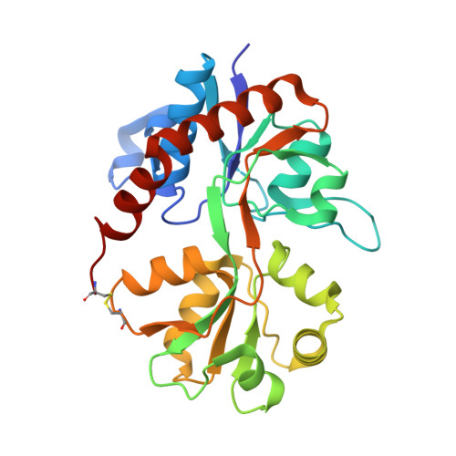
    Two X-ray structures of the GluR2 ligand-binding core in complex with (S)-2-amino-3-(5-tert-butyl-3-hydroxy-4-isoxazolyl)propionic acid ((S)-ATPA) have been determined with and without Zn(2+) ions. (S)-ATPA induces a domain closure of ca. 21 degrees compared to the apo form. The tert-butyl moiety of (S)-ATPA is buried in a partially hydrophobic pocket and forces the ligand into the glutamate-like binding mode. The structures provide new insight into the molecular basis of agonist selectivity between AMPA and kainate receptors.


  • Organizational Affiliation

    Department of Medicinal Chemistry, Pharmaceutical University of Denmark, Universitetsparken 2, DK-2100 Copenhagen, Denmark.


Macromolecules
Find similar proteins by:  (by identity cutoff)  |  3D Structure
Entity ID: 1
MoleculeChains Sequence LengthOrganismDetailsImage
Glutamate receptor 2263Rattus norvegicusMutation(s): 0 
UniProt
Find proteins for P19491 (Rattus norvegicus)
Explore P19491 
Go to UniProtKB:  P19491
Entity Groups  
Sequence Clusters30% Identity50% Identity70% Identity90% Identity95% Identity100% Identity
UniProt GroupP19491
Sequence Annotations
Expand
  • Reference Sequence
Small Molecules
Binding Affinity Annotations 
IDSourceBinding Affinity
CE2 Binding MOAD:  1NNK IC50: 3500 (nM) from 1 assay(s)
PDBBind:  1NNK IC50: 3500 (nM) from 1 assay(s)
Experimental Data & Validation

Experimental Data

  • Method: X-RAY DIFFRACTION
  • Resolution: 1.85 Å
  • R-Value Free: 0.245 
  • R-Value Work: 0.207 
  • Space Group: P 21 21 2
Unit Cell:
Length ( Å )Angle ( ˚ )
a = 54.258α = 90
b = 111.229β = 90
c = 46.643γ = 90
Software Package:
Software NamePurpose
DENZOdata reduction
SCALEPACKdata scaling
AMoREphasing
CNSrefinement

Structure Validation

View Full Validation Report


Ligand Structure Quality Assessment 


Entry History 

Deposition Data

Revision History  (Full details and data files)

  • Version 1.0: 2003-03-04
    Type: Initial release
  • Version 1.1: 2008-04-29
    Changes: Version format compliance
  • Version 1.2: 2011-07-13
    Changes: Derived calculations, Version format compliance
  • Version 1.3: 2017-08-16
    Changes: Data collection, Refinement description, Source and taxonomy
  • Version 1.4: 2024-04-03
    Changes: Data collection, Database references, Derived calculations, Refinement description, Structure summary