1NML

Di-haemic Cytochrome c Peroxidase from Pseudomonas nautica 617, form IN (pH 4.0)


Experimental Data Snapshot

  • Method: X-RAY DIFFRACTION
  • Resolution: 2.20 Å
  • R-Value Free: 0.199 
  • R-Value Work: 0.179 
  • R-Value Observed: 0.179 

wwPDB Validation   3D Report Full Report


Ligand Structure Quality Assessment 


This is version 2.1 of the entry. See complete history


Literature

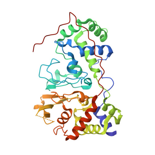
Structural basis for the mechanism of Ca(2+) activation of the di-heme cytochrome c peroxidase from Pseudomonas nautica 617

Dias, J.M.Alves, T.Pereira, A.S.Bourgeois, D.Moura, I.

(2004) Structure 12: 961-973

  • DOI: https://doi.org/10.1016/j.str.2004.03.025
  • Primary Citation of Related Structures:  
    1NML, 1RZ5, 1RZ6

  • PubMed Abstract: 

    Cytochrome c peroxidase (CCP) catalyses the reduction of H(2)O(2) to H(2)O, an important step in the cellular detoxification process. The crystal structure of the di-heme CCP from Pseudomonas nautica 617 was obtained in two different conformations in a redox state with the electron transfer heme reduced. Form IN, obtained at pH 4.0, does not contain Ca(2+) and was refined at 2.2 A resolution. This inactive form presents a closed conformation where the peroxidatic heme adopts a six-ligand coordination, hindering the peroxidatic reaction from taking place. Form OUT is Ca(2+) dependent and was crystallized at pH 5.3 and refined at 2.4 A resolution. This active form shows an open conformation, with release of the distal histidine (His71) ligand, providing peroxide access to the active site. This is the first time that the active and inactive states are reported for a di-heme peroxidase.


  • Organizational Affiliation

    REQUIMTE/CQFB, Departamento de Química, FCT, Universidade Nova de Lisboa, 2829-516 Caparica, Portugal.


Macromolecules
Find similar proteins by:  (by identity cutoff)  |  3D Structure
Entity ID: 1
MoleculeChains Sequence LengthOrganismDetailsImage
di-haem cytochrome c peroxidase326Marinobacter nauticusMutation(s): 0 
EC: 1.11.1.5
UniProt
Find proteins for P83787 (Marinobacter nauticus)
Explore P83787 
Go to UniProtKB:  P83787
Entity Groups  
Sequence Clusters30% Identity50% Identity70% Identity90% Identity95% Identity100% Identity
UniProt GroupP83787
Sequence Annotations
Expand
  • Reference Sequence
Experimental Data & Validation

Experimental Data

  • Method: X-RAY DIFFRACTION
  • Resolution: 2.20 Å
  • R-Value Free: 0.199 
  • R-Value Work: 0.179 
  • R-Value Observed: 0.179 
  • Space Group: P 31 2 1
Unit Cell:
Length ( Å )Angle ( ˚ )
a = 114.457α = 90
b = 114.457β = 90
c = 90.658γ = 120
Software Package:
Software NamePurpose
DENZOdata reduction
SCALEPACKdata scaling
EPMRphasing
CNSrefinement

Structure Validation

View Full Validation Report


Ligand Structure Quality Assessment 


Entry History 

Deposition Data

Revision History  (Full details and data files)

  • Version 1.0: 2004-01-13
    Type: Initial release
  • Version 1.1: 2008-04-29
    Changes: Version format compliance
  • Version 1.2: 2011-07-13
    Changes: Derived calculations, Version format compliance
  • Version 2.0: 2021-03-03
    Changes: Advisory, Atomic model, Data collection, Derived calculations, Non-polymer description, Structure summary
  • Version 2.1: 2023-08-16
    Changes: Data collection, Database references, Refinement description