The mannose-specific bulb lectin from Galanthus nivalis (snowdrop) binds mono- and dimannosides at distinct sites. Structure analysis of refined complexes at 2.3 A and 3.0 A resolution.
Hester, G., Wright, C.S.(1996) J Mol Biol 262: 516-531
- PubMed: 8893860
- DOI: https://doi.org/10.1006/jmbi.1996.0532
- Primary Citation of Related Structures:
1NIV - PubMed Abstract:
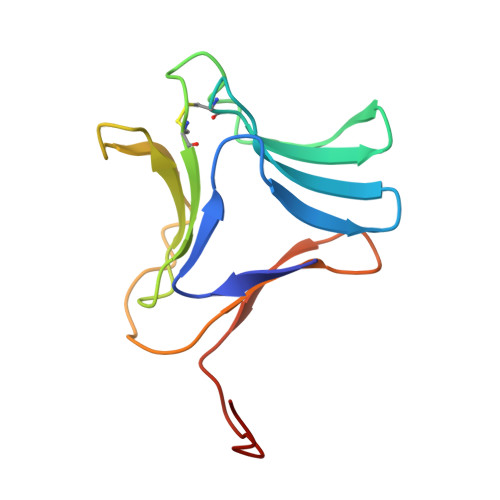
Galanthus nivalis agglutinin (GNA, a 50 kDa tetramer) is a mannose-specific lectin of the Amaryllidaceae family of bulb lectins. Crystal structures of GNA complexed with methyl-alpha-D-mannose (MeMan) and mannose-alpha 1,3-D-mannose-alpha-OMe (MeMan-2) have been determined and analyzed in terms of internal structural symmetry and saccharide binding. The final model of the 2.29 A orthorhombic methyl-alpha-Man complex refined with an R-factor of 0.167 (all data) includes 12 bound sugar ligands and 327 water molecules. The four independent subunits (A, B, C and D) of the 222 tetramer and the three four-stranded beta-sheets (I,II and III) that constitute each subunit compare closely (r.m.s. delta = < 1.0 A). The 12 bound methyl-alpha-Man molecules refined with B-factors < 22 A2 and occupancies in the range of 0.5 to 1.0. The highest occupied site is located in beta-sheet I (site 1), where interactions from the dimer-related subunit contribute to complex stabilization. These subunit pairs (A-D and B-C) associate tightly with a buried surface area of 1738 A2 and 33 interchain hydrogen bonds resulting from C-terminal strand exchange. In comparison, the A-B and C-D subunit pairs have narrow interfaces (476 A2) and no direct H-bond contacts. The 3.0 A structure of the cubic Man-alpha 1,3-Man-OMe complex, determined by molecular replacement and refined with X-PLOR using NCS constraints and density modification methods, is less well ordered due to a high crystal solvent content (68%). Complexed disaccharide is responsible for the most crucial lattice contacts, which involve only one of the two independent subunits (A). The second subunit (C) shows a high degree of flexibility (Bav = 41.7 A2). The complete disaccharide molecule is visible in both subunits at site 3, which is the only extended site. The ligand is oriented with its reducing end positioned in the specificity pocket. The non-reducing manose is in contact through hydrogen bonding with a charged subsite (D37-K38) on the 2-fold-related subunit (A-B or C-D interfaces). Bound Man-alpha 1,3-MeMan is also well defined in site 2 of subunit A, as a result of favorable lattice contacts, while only the mannose residue bound in the specificity pocket is visible at site 2 of subunit C and site 1 of both subunits. Together these results suggest that strong binding correlates with the presence of subsidiary contacts coming either from a dimer-related subunit or from lattice interactions. Site 1 is most specific for terminal non-reducing or reducing mannose, while site 3 is extended and complementary to alpha-1,3 linked mannose oligosaccharides.
Organizational Affiliation:
Department of Biochemistry and Molecular Biophysics, Virginia Commonwealth University, Richmond 23298, USA.