1NDH

CRYSTAL STRUCTURE OF NADH-CYTOCHROME B5 REDUCTASE FROM PIG LIVER AT 2.4 ANGSTROMS RESOLUTION


Experimental Data Snapshot

  • Method: X-RAY DIFFRACTION
  • Resolution: 2.10 Å
  • R-Value Work: 0.223 
  • R-Value Observed: 0.223 

wwPDB Validation   3D Report Full Report


This is version 1.5 of the entry. See complete history


Literature

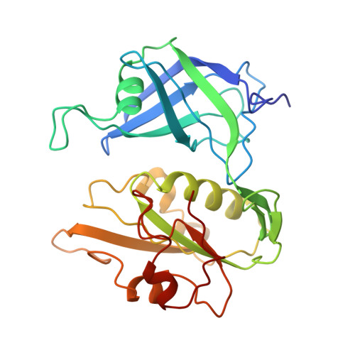
Crystal structure of NADH-cytochrome b5 reductase from pig liver at 2.4 A resolution.

Nishida, H.Inaka, K.Yamanaka, M.Kaida, S.Kobayashi, K.Miki, K.

(1995) Biochemistry 34: 2763-2767

  • DOI: https://doi.org/10.1021/bi00009a004
  • Primary Citation of Related Structures:  
    1NDH

  • PubMed Abstract: 

    The three-dimensional structure of NADH-cytochrome b5 reductase from pig liver microsomes has been determined at 2.4 A resolution by X-ray crystallography. The molecular structure reveals two domains, the FAD binding domain and the NADH domain. A large cleft lies between these two domains and contains the binding site for the FAD prosthetic group. The backbone structure of the FAD binding domain has a great similarity to that of ferredoxin-NADP+ reductase [Karplus, P. A., Daniels, M. J., & Herriott, J. R. (1991) Science 251, 60-65], in spite of the relatively low sequence homology (about 15%) between the two enzymes. On the other hand, the structure of the NADH domain has several structural differences from that of the NADP+ domain of ferredoxin-NADP+ reductase. The size of the cleft between the two domains is larger in NADH-cytochrome b5 reductase than in ferredoxin-NADP+ reductase, which may be responsible for the observed difference in the nucleotide accessibility in the two enzymes.


  • Organizational Affiliation

    Research Laboratory of Resources Utilization, Tokyo Institute of Technology, Yokohama, Japan.


Macromolecules
Find similar proteins by:  (by identity cutoff)  |  3D Structure
Entity ID: 1
MoleculeChains Sequence LengthOrganismDetailsImage
CYTOCHROME B5 REDUCTASE272Sus scrofaMutation(s): 0 
EC: 1.6.2.2
UniProt
Find proteins for P83686 (Sus scrofa)
Explore P83686 
Go to UniProtKB:  P83686
Entity Groups  
Sequence Clusters30% Identity50% Identity70% Identity90% Identity95% Identity100% Identity
UniProt GroupP83686
Sequence Annotations
Expand
  • Reference Sequence
Small Molecules
Ligands 1 Unique
IDChains Name / Formula / InChI Key2D Diagram3D Interactions
FAD
Query on FAD

Download Ideal Coordinates CCD File 
B [auth A]FLAVIN-ADENINE DINUCLEOTIDE
C27 H33 N9 O15 P2
VWWQXMAJTJZDQX-UYBVJOGSSA-N
Experimental Data & Validation

Experimental Data

  • Method: X-RAY DIFFRACTION
  • Resolution: 2.10 Å
  • R-Value Work: 0.223 
  • R-Value Observed: 0.223 
  • Space Group: P 21 21 21
Unit Cell:
Length ( Å )Angle ( ˚ )
a = 86.9α = 90
b = 73.1β = 90
c = 48.9γ = 90
Software Package:
Software NamePurpose
X-PLORmodel building
PROLSQrefinement
X-PLORrefinement
X-PLORphasing

Structure Validation

View Full Validation Report


Entry History 

Deposition Data

Revision History  (Full details and data files)

  • Version 1.0: 1995-02-14
    Type: Initial release
  • Version 1.1: 2008-03-24
    Changes: Version format compliance
  • Version 1.2: 2011-07-13
    Changes: Version format compliance
  • Version 1.3: 2019-07-17
    Changes: Data collection, Other, Refinement description
  • Version 1.4: 2019-08-14
    Changes: Data collection, Refinement description
  • Version 1.5: 2024-02-14
    Changes: Data collection, Database references, Derived calculations