1NCB

CRYSTAL STRUCTURES OF TWO MUTANT NEURAMINIDASE-ANTIBODY COMPLEXES WITH AMINO ACID SUBSTITUTIONS IN THE INTERFACE


Experimental Data Snapshot

  • Method: X-RAY DIFFRACTION
  • Resolution: 2.50 Å
  • R-Value Work: 0.165 
  • R-Value Observed: 0.165 

wwPDB Validation   3D Report Full Report


Ligand Structure Quality Assessment 


This is version 2.0 of the entry. See complete history


Literature

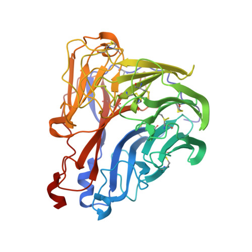
Crystal structures of two mutant neuraminidase-antibody complexes with amino acid substitutions in the interface.

Tulip, W.R.Varghese, J.N.Webster, R.G.Laver, W.G.Colman, P.M.

(1992) J Mol Biol 227: 149-159

  • DOI: https://doi.org/10.1016/0022-2836(92)90688-g
  • Primary Citation of Related Structures:  
    1NCB, 1NCC

  • PubMed Abstract: 

    The site on influenza virus N9 neuraminidase recognized by NC41 monoclonal antibody comprises 19 amino acid residues that are in direct contact with 17 residues on the antibody. Single sequence changes in some of the neuraminidase residues in the site markedly reduce antibody binding. However, two mutants have been found within the site, Ile368 to Arg and Asn329 to Asp selected by antibodies other than NC41, and these mutants bind NC41 antibody with only slightly reduced affinity. The three-dimensional structures of the two mutant N9-NC41 antibody complexes as derived from the wild-type complex are presented. Both structures show that some amino acid substitutions can be accommodated within an antigen-antibody interface by local structural rearrangements around the mutation site. In the Ile368 to Arg mutant complex, the side-chain of Arg368 is shifted by 2.9 A from its position in the uncomplexed mutant and a shift of 1.3 A in the position of the light chain residue HisL55 with respect to the wild-type complex is also observed. In the other mutant, the side-chain of Asp329 appears rotated by 150 degrees around C alpha-C beta with respect to the uncomplexed mutant, so that the carboxylate group is moved to the periphery of the antigen-antibody interface. The results provide a basis for understanding some of the potential structural effects of somatic hypermutation on antigen-antibody binding in those cases where the mutation in the antibody occurs at antigen-contacting residues, and demonstrate again the importance of structural context in evaluating the effect of amino acid substitutions on protein structure and function.


  • Organizational Affiliation

    CSIRO Division of Biomolecular Engineering, Parkville, Victoria, Australia.


Macromolecules
Find similar proteins by:  (by identity cutoff)  |  3D Structure
Entity ID: 1
MoleculeChains Sequence LengthOrganismDetailsImage
INFLUENZA A SUBTYPE N9 NEURAMINIDASEA [auth N]389Influenza A virus (A/tern/Australia/G70C/1975(H11N9))Mutation(s): 0 
EC: 3.2.1.18
UniProt
Find proteins for P03472 (Influenza A virus (strain A/Tern/Australia/G70C/1975 H11N9))
Explore P03472 
Go to UniProtKB:  P03472
Entity Groups  
Sequence Clusters30% Identity50% Identity70% Identity90% Identity95% Identity100% Identity
UniProt GroupP03472
Sequence Annotations
Expand
  • Reference Sequence
Find similar proteins by:  (by identity cutoff)  |  3D Structure
Entity ID: 2
MoleculeChains Sequence LengthOrganismDetailsImage
IGG2A-KAPPA NC41 FAB (LIGHT CHAIN)B [auth L]214Mus musculusMutation(s): 0 
Entity Groups  
Sequence Clusters30% Identity50% Identity70% Identity90% Identity95% Identity100% Identity
Sequence Annotations
Expand
  • Reference Sequence
Find similar proteins by:  (by identity cutoff)  |  3D Structure
Entity ID: 3
MoleculeChains Sequence LengthOrganismDetailsImage
IGG2A-KAPPA NC41 FAB (HEAVY CHAIN)C [auth H]221Mus musculusMutation(s): 0 
UniProt
Find proteins for P01865 (Mus musculus)
Explore P01865 
Go to UniProtKB:  P01865
Entity Groups  
Sequence Clusters30% Identity50% Identity70% Identity90% Identity95% Identity100% Identity
UniProt GroupP01865
Sequence Annotations
Expand
  • Reference Sequence
Oligosaccharides

Help

Entity ID: 4
MoleculeChains Length2D Diagram Glycosylation3D Interactions
alpha-D-mannopyranose-(1-2)-alpha-D-mannopyranose-(1-2)-alpha-D-mannopyranose-(1-3)-beta-D-mannopyranose-(1-4)-2-acetamido-2-deoxy-beta-D-glucopyranose-(1-4)-2-acetamido-2-deoxy-beta-D-glucopyranoseD [auth A]6N-Glycosylation
Glycosylation Resources
GlyTouCan:  G01760ZU
GlyCosmos:  G01760ZU
GlyGen:  G01760ZU
Entity ID: 5
MoleculeChains Length2D Diagram Glycosylation3D Interactions
2-acetamido-2-deoxy-beta-D-glucopyranose-(1-4)-2-acetamido-2-deoxy-beta-D-glucopyranoseE [auth B]2N-Glycosylation
Glycosylation Resources
GlyTouCan:  G42666HT
GlyCosmos:  G42666HT
GlyGen:  G42666HT
Small Molecules
Ligands 2 Unique
IDChains Name / Formula / InChI Key2D Diagram3D Interactions
NAG
Query on NAG

Download Ideal Coordinates CCD File 
F [auth N]2-acetamido-2-deoxy-beta-D-glucopyranose
C8 H15 N O6
OVRNDRQMDRJTHS-FMDGEEDCSA-N
CA
Query on CA

Download Ideal Coordinates CCD File 
G [auth N]CALCIUM ION
Ca
BHPQYMZQTOCNFJ-UHFFFAOYSA-N
Experimental Data & Validation

Experimental Data

  • Method: X-RAY DIFFRACTION
  • Resolution: 2.50 Å
  • R-Value Work: 0.165 
  • R-Value Observed: 0.165 
  • Space Group: P 4 21 2
Unit Cell:
Length ( Å )Angle ( ˚ )
a = 167α = 90
b = 167β = 90
c = 124γ = 90
Software Package:
Software NamePurpose
X-PLORmodel building
X-PLORrefinement
X-PLORphasing

Structure Validation

View Full Validation Report


Ligand Structure Quality Assessment 


Entry History 

Deposition Data

Revision History  (Full details and data files)

  • Version 1.0: 1994-01-31
    Type: Initial release
  • Version 1.1: 2008-03-24
    Changes: Version format compliance
  • Version 1.2: 2011-07-13
    Changes: Derived calculations, Source and taxonomy, Version format compliance
  • Version 2.0: 2020-07-29
    Type: Remediation
    Reason: Carbohydrate remediation
    Changes: Atomic model, Data collection, Database references, Derived calculations, Other, Structure summary