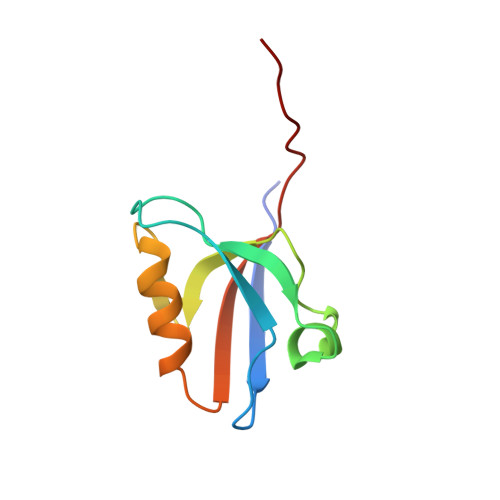
Crystal structure of GRIP1 PDZ6-peptide complex reveals the structural basis for class II PDZ target recognition and PDZ domain-mediated multimerization
Im, Y.J., Park, S.H., Rho, S.H., Lee, J.H., Kang, G.B., Sheng, M., Kim, E., Eom, S.H.(2003) J Biol Chem 278: 8501-8507
- PubMed: 12493751
- DOI: https://doi.org/10.1074/jbc.M212263200
- Primary Citation of Related Structures:
1N7E, 1N7F - PubMed Abstract:
PDZ domains bind to short segments within target proteins in a sequence-specific fashion. Glutamate receptor-interacting protein (GRIP)/ABP family proteins contain six to seven PDZ domains and interact via the sixth PDZ domain (class II) with the C termini of various proteins including liprin-alpha. In addition the PDZ456 domain mediates the formation of homo- and heteromultimers of GRIP proteins. To better understand the structural basis of peptide recognition by a class II PDZ domain and PDZ-mediated multimerization, we determined the crystal structures of the GRIP1 PDZ6 domain alone and in complex with a synthetic C-terminal octapeptide of human liprin-alpha at resolutions of 1.5 and 1.8 A, respectively. Remarkably, unlike other class II PDZ domains, Ile-736 at alphaB5 rather than conserved Leu-732 at alphaB1 makes a direct hydrophobic contact with the side chain of the Tyr at the -2 position of the ligand. Moreover, the peptide-bound structure of PDZ6 shows a slight reorientation of helix alphaB, indicating that the second hydrophobic pocket undergoes a conformational adaptation to accommodate the bulkiness of the Tyr side chain, and forms an antiparallel dimer through an interface located at a site distal to the peptide-binding groove. This configuration may enable formation of GRIP multimers and efficient clustering of GRIP-binding proteins.
Organizational Affiliation:
Department of Life Science, Kwangju Institute of Science and Technology, Gwangju 500-712, South Korea.