1N44

Crystal Structure of Annexin V R23E Mutant


Experimental Data Snapshot

  • Method: X-RAY DIFFRACTION
  • Resolution: 3.00 Å
  • R-Value Free: 0.241 
  • R-Value Work: 0.163 
  • R-Value Observed: 0.163 

wwPDB Validation   3D Report Full Report


This is version 1.4 of the entry. See complete history


Literature

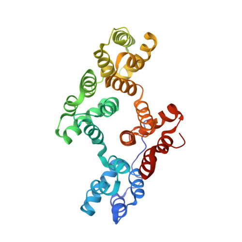
Interfacial basic cluster in anexin V couples phospholipid binding and trimer formation on membrane surfaces

Mo, Y.D.Campos, B.Mealy, T.R.Commodore, L.Head, J.F.Dedman, J.R.Seaton, B.A.

(2003) J Biol Chem 278: 2437-2443

  • DOI: https://doi.org/10.1074/jbc.M210286200
  • Primary Citation of Related Structures:  
    1N41, 1N42, 1N44

  • PubMed Abstract: 

    Annexin V is an abundant eukaryotic protein that binds phospholipid membranes in a Ca(2+)-dependent manner. In the present studies, site-directed mutagenesis was combined with x-ray crystallography and solution liposome binding assays to probe the functional role of a cluster of interfacial basic residues in annexin V. Four mutants were investigated: R23E, K27E, R61E, and R149E. All four mutants exhibited a significant reduction in adsorption to phospholipid membranes relative to the wild-type protein, and the R23E mutation was the most deleterious. Crystal structures of wild-type and mutant proteins were similar except for local changes in salt bridges involving basic cluster residues. The combined data indicate that Arg(23) is a major determinant for interfacial phospholipid binding and participates in an intermolecular salt bridge that is key for trimer formation on the membrane surface. Together, crystallographic and solution data provide evidence that the interfacial basic cluster is a locus where trimerization is synergistically coupled to membrane phospholipid binding.


  • Organizational Affiliation

    Department of Physiology and Biophysics, Boston University School of Medicine, Massachusetts 02118, USA.


Macromolecules
Find similar proteins by:  (by identity cutoff)  |  3D Structure
Entity ID: 1
MoleculeChains Sequence LengthOrganismDetailsImage
Annexin V319Rattus norvegicusMutation(s): 1 
UniProt
Find proteins for P14668 (Rattus norvegicus)
Explore P14668 
Go to UniProtKB:  P14668
Entity Groups  
Sequence Clusters30% Identity50% Identity70% Identity90% Identity95% Identity100% Identity
UniProt GroupP14668
Sequence Annotations
Expand
  • Reference Sequence
Experimental Data & Validation

Experimental Data

  • Method: X-RAY DIFFRACTION
  • Resolution: 3.00 Å
  • R-Value Free: 0.241 
  • R-Value Work: 0.163 
  • R-Value Observed: 0.163 
  • Space Group: H 3
Unit Cell:
Length ( Å )Angle ( ˚ )
a = 157.757α = 90
b = 157.757β = 90
c = 36.761γ = 120
Software Package:
Software NamePurpose
DENZOdata reduction
SCALEPACKdata scaling
CNSrefinement
CNSphasing

Structure Validation

View Full Validation Report


Entry History 

Deposition Data

Revision History  (Full details and data files)

  • Version 1.0: 2003-02-04
    Type: Initial release
  • Version 1.1: 2008-04-28
    Changes: Version format compliance
  • Version 1.2: 2011-07-13
    Changes: Version format compliance
  • Version 1.3: 2021-10-27
    Changes: Database references, Derived calculations
  • Version 1.4: 2024-02-14
    Changes: Data collection