1N3N

Crystal structure of a mycobacterial hsp60 epitope with the murine class I MHC molecule H-2Db


Experimental Data Snapshot

  • Method: X-RAY DIFFRACTION
  • Resolution: 3.00 Å
  • R-Value Free: 0.307 
  • R-Value Work: 0.246 

wwPDB Validation   3D Report Full Report


This is version 1.2 of the entry. See complete history


Literature

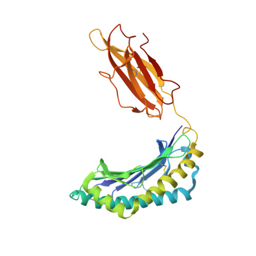
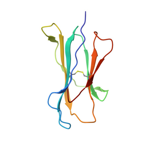
Structural analysis of mycobacterial and murine hsp60 epitopes in complex with the class I MHC molecule H-2D(b)

Ciatto, C.Capitani, G.Tissot, A.C.Pecorari, F.Plueckthun, A.Gruetter, M.G.

(2003) FEBS Lett 543: 11-15

  • DOI: https://doi.org/10.1016/s0014-5793(03)00325-9
  • Primary Citation of Related Structures:  
    1N3N

  • PubMed Abstract: 

    The decameric peptide SALQNAASIA from the Mycobacterium bovis heat shock protein (hsp) 60 is recognized by the murine T-cell receptor UZ-3-4 in complex with the murine class I major histocompatibility complex molecule H-2D(b). This T-cell receptor cross-reacts with the H-2D(b)-bound non-homologous decameric peptide KDIGNIISDA from the murine hsp60, but does not recognize the nonameric mycobacterial peptide SALQNAASI. Cross-recognition of the KDIGNIISDA/H-2D(b) complex induces autoimmune pathology in immunodeficient mice. We solved the X-ray crystal structure of the SALQNAASIA/H-2D(b) complex at 3.0 A resolution, and we modelled the KDIGNIISDA and SALQNAASI peptides in the H-2D(b) binding site. The structural analysis of the H-2D(b)-bound hsp60 epitopes offers insight into T-cell receptor cross-reactivity.


  • Organizational Affiliation

    Department of Biochemistry, University of Zürich, Winterthurerstrasse 190, CH-8057, Zürich, Switzerland.


Macromolecules
Find similar proteins by:  (by identity cutoff)  |  3D Structure
Entity ID: 1
MoleculeChains Sequence LengthOrganismDetailsImage
H-2 CLASS I HISTOCOMPATIBILITY ANTIGEN, D-B ALPHA CHAIN
A, C, E, G
280Mus musculusMutation(s): 0 
UniProt
Find proteins for P01899 (Mus musculus)
Explore P01899 
Go to UniProtKB:  P01899
Entity Groups  
Sequence Clusters30% Identity50% Identity70% Identity90% Identity95% Identity100% Identity
UniProt GroupP01899
Sequence Annotations
Expand
  • Reference Sequence
Find similar proteins by:  (by identity cutoff)  |  3D Structure
Entity ID: 2
MoleculeChains Sequence LengthOrganismDetailsImage
BETA-2-MICROGLOBULIN
B, D, F, H
99Mus musculusMutation(s): 0 
UniProt & NIH Common Fund Data Resources
Find proteins for P01887 (Mus musculus)
Explore P01887 
Go to UniProtKB:  P01887
IMPC:  MGI:88127
Entity Groups  
Sequence Clusters30% Identity50% Identity70% Identity90% Identity95% Identity100% Identity
UniProt GroupP01887
Sequence Annotations
Expand
  • Reference Sequence

Find similar proteins by:  Sequence   |   3D Structure  

Entity ID: 3
MoleculeChains Sequence LengthOrganismDetailsImage
mycobacterial hsp60 decameric epitope
I, J, K, L
10N/AMutation(s): 0 
Entity Groups  
Sequence Clusters30% Identity50% Identity70% Identity90% Identity95% Identity100% Identity
Sequence Annotations
Expand
  • Reference Sequence
Experimental Data & Validation

Experimental Data

  • Method: X-RAY DIFFRACTION
  • Resolution: 3.00 Å
  • R-Value Free: 0.307 
  • R-Value Work: 0.246 
  • Space Group: C 1 2 1
Unit Cell:
Length ( Å )Angle ( ˚ )
a = 129.276α = 90
b = 122.995β = 90.01
c = 150.833γ = 90
Software Package:
Software NamePurpose
DENZOdata reduction
SCALEPACKdata scaling
AMoREphasing
CNSrefinement

Structure Validation

View Full Validation Report


Entry History 

Deposition Data

Revision History  (Full details and data files)

  • Version 1.0: 2003-05-27
    Type: Initial release
  • Version 1.1: 2008-04-28
    Changes: Version format compliance
  • Version 1.2: 2011-07-13
    Changes: Version format compliance