1N2A

Crystal Structure of a Bacterial Glutathione Transferase from Escherichia coli with Glutathione Sulfonate in the Active Site


Experimental Data Snapshot

  • Method: X-RAY DIFFRACTION
  • Resolution: 1.90 Å
  • R-Value Free: 0.260 
  • R-Value Work: 0.195 
  • R-Value Observed: 0.201 

wwPDB Validation   3D Report Full Report


Ligand Structure Quality Assessment 


This is version 1.3 of the entry. See complete history


Literature

Conserved structural elements in glutathione transferase homologues encoded in the genome of Escherichia coli

Rife, C.L.Parsons, J.F.Xiao, G.Gilliland, G.L.Armstrong, R.N.

(2003) Proteins 53: 777-782

  • DOI: https://doi.org/10.1002/prot.10452
  • Primary Citation of Related Structures:  
    1N2A

  • PubMed Abstract: 

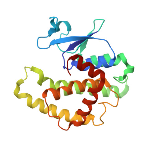
    Multiple sequence alignments of the eight glutathione (GSH) transferase homologues encoded in the genome of Escherichia coli were used to define a consensus sequence for the proteins. The consensus sequence was analyzed in the context of the three-dimensional structure of the gst gene product (EGST) obtained from two different crystal forms of the enzyme. The enzyme consists of two domains. The N-terminal region (domain I) has a thioredoxin-like alpha/beta-fold, while the C-terminal domain (domain II) is all alpha-helical. The majority of the consensus residues (12/17) reside in the N-terminal domain. Fifteen of the 17 residues are involved in hydrophobic core interactions, turns, or electrostatic interactions between the two domains. The results suggest that all of the homologues retain a well-defined group of structural elements both in and between the N-terminal alpha/beta domain and the C-terminal domain. The conservation of two key residues for the recognition motif for the gamma-glutamyl-portion of GSH indicates that the homologues may interact with GSH or GSH analogues such as glutathionylspermidine or alpha-amino acids. The genome context of two of the homologues forms the basis for a hypothesis that the b2989 and yibF gene products are involved in glutathionylspermidine and selenium biochemistry, respectively.


  • Organizational Affiliation

    Department of Biochemistry and the Center in Molecular Toxicology, Vanderbilt University School of Medicine, Nashville Tennessee 37232-0146, USA.


Macromolecules
Find similar proteins by:  (by identity cutoff)  |  3D Structure
Entity ID: 1
MoleculeChains Sequence LengthOrganismDetailsImage
Glutathione S-transferase
A, B
201Escherichia coliMutation(s): 0 
Gene Names: GT_1787923
EC: 2.5.1.18
UniProt
Find proteins for P0A9D2 (Escherichia coli (strain K12))
Explore P0A9D2 
Go to UniProtKB:  P0A9D2
Entity Groups  
Sequence Clusters30% Identity50% Identity70% Identity90% Identity95% Identity100% Identity
UniProt GroupP0A9D2
Sequence Annotations
Expand
  • Reference Sequence
Small Molecules
Experimental Data & Validation

Experimental Data

  • Method: X-RAY DIFFRACTION
  • Resolution: 1.90 Å
  • R-Value Free: 0.260 
  • R-Value Work: 0.195 
  • R-Value Observed: 0.201 
  • Space Group: P 21 21 21
Unit Cell:
Length ( Å )Angle ( ˚ )
a = 47.616α = 90
b = 57.166β = 90
c = 134.927γ = 90
Software Package:
Software NamePurpose
DENZOdata reduction
SCALEPACKdata scaling
AMoREphasing
REFMACrefinement

Structure Validation

View Full Validation Report


Ligand Structure Quality Assessment 


Entry History 

Deposition Data

Revision History  (Full details and data files)

  • Version 1.0: 2003-11-04
    Type: Initial release
  • Version 1.1: 2008-04-28
    Changes: Version format compliance
  • Version 1.2: 2011-07-13
    Changes: Version format compliance
  • Version 1.3: 2024-02-14
    Changes: Data collection, Database references, Derived calculations, Refinement description