The high resolution structures of free and inhibitor-bound Trypanosoma rangeli sialidase and its comparison with T. cruzi trans-sialidase
Amaya, M.F., Buschiazzo, A., Nguyen, T., Alzari, P.M.(2003) J Mol Biol 325: 773-784
- PubMed: 12507479
- DOI: https://doi.org/10.1016/s0022-2836(02)01306-2
- Primary Citation of Related Structures:
1N1S, 1N1T, 1N1V, 1N1Y - PubMed Abstract:
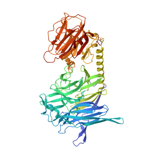
The structure of the recombinant Trypanosoma rangeli sialidase (TrSA) has been determined at 1.6A resolution, and the structures of its complexes with the transition state analog inhibitor 2-deoxy-2,3-dehydro-N-acetyl-neuraminic acid (DANA), Neu-5-Ac-thio-alpha(2,3)-galactoside (NATG) and N-acetylneuraminic acid (NANA) have been determined at 1.64A, 2.1A and 2.85A, respectively. The 3D structure of TrSA is essentially identical to that of the natural enzyme, except for the absence of covalently attached sugar at five distinct N-glycosylation sites. The protein exhibits a topologically rigid active site architecture that is unaffected by ligand binding. The overall binding of DANA to the active site cleft is similar to that observed for other viral and bacterial sialidases, dominated by the interactions of the inhibitor carboxylate with the conserved arginine triad. However, the interactions of the other pyranoside ring substituents (hydroxyl, N-acetyl and glycerol moieties) differ between trypanosomal, bacterial and viral sialidases, providing a structural basis for specific inhibitor design. Sialic acid is found to bind the enzyme with the sugar ring in a distorted (half-chair or boat) conformation and the 2-OH hydroxyl group at hydrogen bonding distance of the carboxylate of Asp60, substantiating a direct catalytic role for this residue. A detailed comparison of TrSA with the closely related structure of T.cruzi trans-sialidase (TcTS) reveals a highly conserved catalytic center, where subtle structural differences account for strikingly different enzymatic activities and inhibition properties. The structure of TrSA in complex with NATG shows the active site cleft occupied by a smaller compound which could be identified as DANA, probably the product of a hydrolytic side reaction. Indeed, TrSA (but not TcTS) was found to cleave O and S-linked sialylated substrates, further stressing the functional differences between trypanosomal sialidases and trans-sialidases.
Organizational Affiliation:
Unité de Biochimie Structurale, CNRS URA 2185, Institut Pasteur, 25 rue du Dr. Roux, 75724 Paris cédex 15, France.