1N0J

The Structure of Human Mitochondrial MN3+ Superoxide Dismutase Reveals a Novel Tetrameric Interface of Two 4-Helix Bundles


Experimental Data Snapshot

  • Method: X-RAY DIFFRACTION
  • Resolution: 2.20 Å
  • R-Value Work: 0.171 
  • R-Value Observed: 0.171 

wwPDB Validation   3D Report Full Report


This is version 1.4 of the entry. See complete history


Literature

The Structure of Human Mitochondrial Mn3+ Superoxide Dismutase Reveals a Novel Tetrameric Interface of Two 4-Helix Bundles

Borgstahl, G.E.Parge, H.E.Hickey, M.J.Beyer Jr., W.F.Hallewell, R.A.Tainer, J.A.

(1992) Cell 71: 107-107

  • DOI: https://doi.org/10.1016/0092-8674(92)90270-m
  • Primary Citation of Related Structures:  
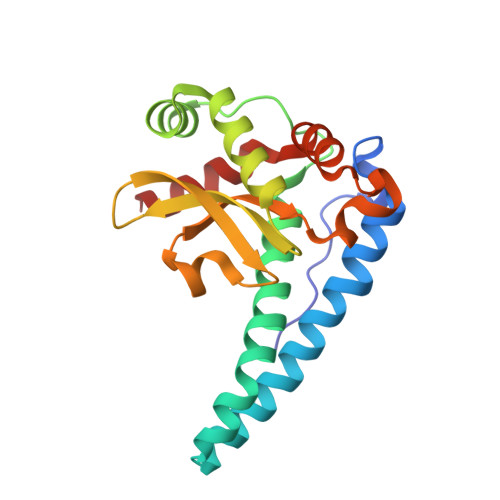
    1N0J

  • PubMed Abstract: 

    The 2.2 A resolution crystal structure of recombinant human manganese superoxide dismutase, a homotetrameric enzyme that protects mitochondria against oxygen-mediated free radical damage, has been determined. Within each subunit, both the N-terminal helical hairpin and C-terminal alpha/beta domains contribute ligands to the catalytic manganese site. Two identical 4-helix bundles, symmetrically assembled from the N-terminal helical hairpins, form novel tetrameric interfaces that stabilize the active sites. Structurally altered polymorphic variants with reduced activity, such as tetrameric interface mutant Ile-58 to Thr, may produce not only an early selective advantage, through enhanced cytotoxicity of tumor necrosis factor for virus-infected cells, but also detrimental effects from increased mitochondrial oxidative damage, contributing to degenerative conditions, including diabetes, aging, and Parkinson's and Alzheimer's diseases.


  • Organizational Affiliation

    Department of Molecular Biology, Scripps Research Institute, La Jolla, California 92037.


Macromolecules
Find similar proteins by:  (by identity cutoff)  |  3D Structure
Entity ID: 1
MoleculeChains Sequence LengthOrganismDetailsImage
Superoxide dismutase [Mn]
A, B
199Homo sapiensMutation(s): 0 
EC: 1.15.1.1
UniProt & NIH Common Fund Data Resources
Find proteins for P04179 (Homo sapiens)
Explore P04179 
Go to UniProtKB:  P04179
PHAROS:  P04179
Entity Groups  
Sequence Clusters30% Identity50% Identity70% Identity90% Identity95% Identity100% Identity
UniProt GroupP04179
Sequence Annotations
Expand
  • Reference Sequence
Small Molecules
Ligands 1 Unique
IDChains Name / Formula / InChI Key2D Diagram3D Interactions
MN
Query on MN

Download Ideal Coordinates CCD File 
C [auth A],
D [auth B]
MANGANESE (II) ION
Mn
WAEMQWOKJMHJLA-UHFFFAOYSA-N
Experimental Data & Validation

Experimental Data

  • Method: X-RAY DIFFRACTION
  • Resolution: 2.20 Å
  • R-Value Work: 0.171 
  • R-Value Observed: 0.171 
  • Space Group: P 21 21 2
Unit Cell:
Length ( Å )Angle ( ˚ )
a = 76.13α = 90
b = 79.9β = 90
c = 68.05γ = 90
Software Package:
Software NamePurpose
X-PLORrefinement
X-GENdata reduction

Structure Validation

View Full Validation Report


Entry History 

Deposition Data

Revision History  (Full details and data files)

  • Version 1.0: 2002-11-06
    Type: Initial release
  • Version 1.1: 2007-10-16
    Changes: Version format compliance
  • Version 1.2: 2011-07-13
    Changes: Derived calculations, Version format compliance
  • Version 1.3: 2017-10-11
    Changes: Derived calculations, Refinement description
  • Version 1.4: 2024-02-14
    Changes: Data collection, Database references, Derived calculations