1MZR

Structure of dkga from E.coli at 2.13 A resolution solved by molecular replacement


Experimental Data Snapshot

  • Method: X-RAY DIFFRACTION
  • Resolution: 2.13 Å
  • R-Value Free: 0.220 
  • R-Value Work: 0.175 

wwPDB Validation   3D Report Full Report


This is version 1.3 of the entry. See complete history


Literature

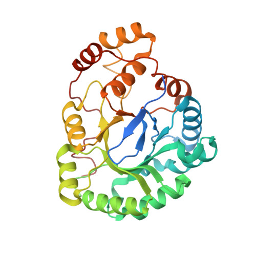
Crystal structure of Escherichia coli DkgA, a broad-specificity aldo-keto reductase.

Jeudy, S.Monchois, V.Maza, C.Claverie, J.M.Abergel, C.

(2006) Proteins 62: 302-307


Macromolecules
Find similar proteins by:  (by identity cutoff)  |  3D Structure
Entity ID: 1
MoleculeChains Sequence LengthOrganismDetailsImage
2,5-diketo-D-gluconate reductase A
A, B
296Escherichia coliMutation(s): 0 
Gene Names: yqhe
EC: 1.1.1
UniProt
Find proteins for Q46857 (Escherichia coli (strain K12))
Explore Q46857 
Go to UniProtKB:  Q46857
Entity Groups  
Sequence Clusters30% Identity50% Identity70% Identity90% Identity95% Identity100% Identity
UniProt GroupQ46857
Sequence Annotations
Expand
  • Reference Sequence
Experimental Data & Validation

Experimental Data

  • Method: X-RAY DIFFRACTION
  • Resolution: 2.13 Å
  • R-Value Free: 0.220 
  • R-Value Work: 0.175 
  • Space Group: C 2 2 21
Unit Cell:
Length ( Å )Angle ( ˚ )
a = 139.177α = 90
b = 145.697β = 90
c = 79.511γ = 90
Software Package:
Software NamePurpose
MOSFLMdata reduction
SCALAdata scaling
AMoREphasing
CNSrefinement
CCP4data scaling

Structure Validation

View Full Validation Report


Entry History 

Deposition Data

Revision History  (Full details and data files)

  • Version 1.0: 2003-10-28
    Type: Initial release
  • Version 1.1: 2008-04-28
    Changes: Version format compliance
  • Version 1.2: 2011-07-13
    Changes: Non-polymer description, Version format compliance
  • Version 1.3: 2023-10-25
    Changes: Data collection, Database references, Derived calculations, Refinement description