1MZJ

Crystal Structure of the Priming beta-Ketosynthase from the R1128 Polyketide Biosynthetic Pathway


Experimental Data Snapshot

  • Method: X-RAY DIFFRACTION
  • Resolution: 2.10 Å
  • R-Value Free: 0.251 
  • R-Value Work: 0.215 
  • R-Value Observed: 0.217 

wwPDB Validation   3D Report Full Report


Ligand Structure Quality Assessment 


This is version 1.2 of the entry. See complete history


Literature

Crystal structure of the priming beta-ketosynthase from the R1128 polyketide biosynthetic pathway

Pan, H.Tsai, S.Meadows, E.S.Miercke, L.J.Keatinge-Clay, A.T.O'Connell, J.Khosla, C.Stroud, R.M.

(null) Structure 10: 1559-1568

  • DOI: https://doi.org/10.1016/s0969-2126(02)00889-4
  • Primary Citation of Related Structures:  
    1MZJ

  • PubMed Abstract: 

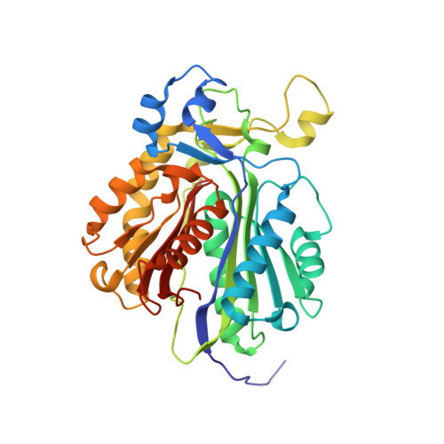
    ZhuH is a priming ketosynthase that initiates the elongation of the polyketide chain in the biosynthetic pathway of a type II polyketide, R1128. The crystal structure of ZhuH in complex with the priming substrate acetyl-CoA reveals an extensive loop region at the dimer interface that appears to affect the selectivity for the primer unit. Acetyl-CoA is bound in a 20 A-long channel, which placed the acetyl group against the catalytic triad. Analysis of the primer unit binding site in ZhuH suggests that it can accommodate acyl chains that are two to four carbons long. Selectivity and primer unit size appear to involve the side chains of three residues on the loops close to the dimer interface that constitute the bottom of the substrate binding pocket.


  • Organizational Affiliation

    Department of Biophysics and Biochemistry, University of California San Francisco, San Francisco, CA 94143, USA.


Macromolecules
Find similar proteins by:  (by identity cutoff)  |  3D Structure
Entity ID: 1
MoleculeChains Sequence LengthOrganismDetailsImage
Beta-ketoacylsynthase III
A, B
339Streptomyces sp. R1128Mutation(s): 0 
Gene Names: zhuH
EC: 2.3.1.41
UniProt
Find proteins for Q9F6D4 (Streptomyces lividans)
Explore Q9F6D4 
Go to UniProtKB:  Q9F6D4
Entity Groups  
Sequence Clusters30% Identity50% Identity70% Identity90% Identity95% Identity100% Identity
UniProt GroupQ9F6D4
Sequence Annotations
Expand
  • Reference Sequence
Experimental Data & Validation

Experimental Data

  • Method: X-RAY DIFFRACTION
  • Resolution: 2.10 Å
  • R-Value Free: 0.251 
  • R-Value Work: 0.215 
  • R-Value Observed: 0.217 
  • Space Group: P 31
Unit Cell:
Length ( Å )Angle ( ˚ )
a = 96α = 90
b = 96β = 90
c = 72.6γ = 120
Software Package:
Software NamePurpose
DENZOdata reduction
SCALEPACKdata scaling
CNSrefinement
CNSphasing

Structure Validation

View Full Validation Report


Ligand Structure Quality Assessment 


Entry History 

Deposition Data

Revision History  (Full details and data files)

  • Version 1.0: 2002-12-11
    Type: Initial release
  • Version 1.1: 2008-04-28
    Changes: Version format compliance
  • Version 1.2: 2011-07-13
    Changes: Version format compliance