1MW9

Crystal Structure of H365R mutant of 67 kDA N-terminal fragment of E. coli DNA Topoisomerase I


Experimental Data Snapshot

  • Method: X-RAY DIFFRACTION
  • Resolution: 1.67 Å
  • R-Value Free: 0.216 
  • R-Value Work: 0.208 
  • R-Value Observed: 0.209 

wwPDB Validation   3D Report Full Report


This is version 1.6 of the entry. See complete history


Literature

Structure of a Complex between E. coli DNA Topoisomerase I and Single-Stranded DNA.

Perry, K.Mondragon, A.

(2003) Structure 11: 1349-1358

  • DOI: https://doi.org/10.1016/j.str.2003.09.013
  • Primary Citation of Related Structures:  
    1MW8, 1MW9

  • PubMed Abstract: 

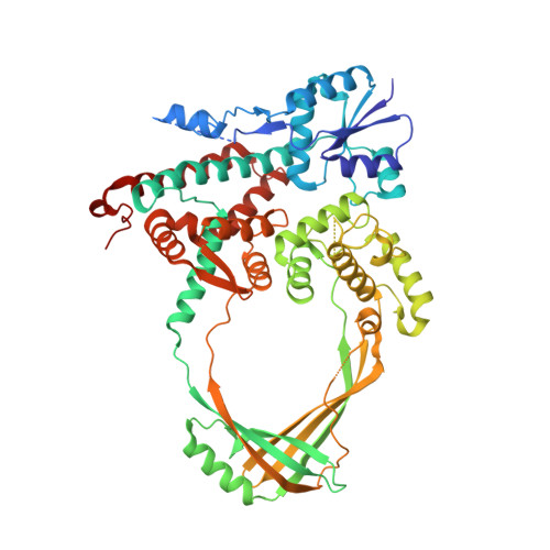
    In order to gain insights into the mechaism of ssDNA binding and recognition by Escherichia coli DNA topoisomerase I, the structure of the 67 kDa N-terminal fragment of topoisomerase I was solved in complex with ssDNA. The structure reveals a new conformational stage in the multistep catalytic cycle of type IA topoisomerases. In the structure, the ssDNA binding groove leading to the active site is occupied, but the active site is not fully formed. Large conformational changes are not seen; instead, a single helix parallel to the ssDNA binding groove shifts to clamp the ssDNA. The structure helps clarify the temporal sequence of conformational events, starting from an initial empty enzyme and proceeding to a ssDNA-occupied and catalytically competent active site.


  • Organizational Affiliation

    Department of Biochemistry, Molecular Biology, and Cell Biology, Northwestern University, 2205 Tech Drive, Evanston, IL 60208, USA.


Macromolecules
Find similar proteins by:  (by identity cutoff)  |  3D Structure
Entity ID: 1
MoleculeChains Sequence LengthOrganismDetailsImage
DNA Topoisomerase IA [auth X]592Escherichia coliMutation(s): 1 
Gene Names: TopA
EC: 5.99.1.2
UniProt
Find proteins for P06612 (Escherichia coli (strain K12))
Explore P06612 
Go to UniProtKB:  P06612
Entity Groups  
Sequence Clusters30% Identity50% Identity70% Identity90% Identity95% Identity100% Identity
UniProt GroupP06612
Sequence Annotations
Expand
  • Reference Sequence
Experimental Data & Validation

Experimental Data

  • Method: X-RAY DIFFRACTION
  • Resolution: 1.67 Å
  • R-Value Free: 0.216 
  • R-Value Work: 0.208 
  • R-Value Observed: 0.209 
  • Space Group: P 21 21 21
Unit Cell:
Length ( Å )Angle ( ˚ )
a = 62.99α = 90
b = 78.705β = 90
c = 139.328γ = 90
Software Package:
Software NamePurpose
REFMACrefinement
SCALAdata scaling
CNSrefinement
MAR345data collection
CCP4data scaling
CNSphasing

Structure Validation

View Full Validation Report


Entry History 

Deposition Data

Revision History  (Full details and data files)

  • Version 1.0: 2003-10-14
    Type: Initial release
  • Version 1.1: 2008-04-28
    Changes: Version format compliance
  • Version 1.2: 2011-07-13
    Changes: Version format compliance
  • Version 1.3: 2017-10-11
    Changes: Refinement description
  • Version 1.4: 2019-07-24
    Changes: Data collection, Refinement description
  • Version 1.5: 2021-10-27
    Changes: Database references, Derived calculations
  • Version 1.6: 2024-02-14
    Changes: Data collection, Refinement description