The Crystal Structure of the Phosphorylation Domain in PhoP Reveals a Functional Tandem Association Mediated by an Asymmetric Interface
Birck, C., Chen, Y., Hulett, F.M., Samama, J.P.(2003) J Bacteriol 185: 254-261
- PubMed: 12486062
- DOI: https://doi.org/10.1128/JB.185.1.254-261.2003
- Primary Citation of Related Structures:
1MVO - PubMed Abstract:
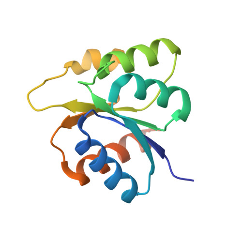
PhoP from Bacillus subtilis belongs to the OmpR subfamily of response regulators. It regulates the transcription of several operons and participates in a signal transduction network that controls adaptation of the bacteria to phosphate deficiency. The receiver domains of two members of this subfamily, PhoB from Escherichia coli and DrrD from Thermotoga maritima, have been structurally characterized. These modules have similar overall folds but display remarkable differences in the conformation of the beta4-alpha4 and alpha4 regions. The crystal structure of the receiver domain of PhoP (PhoPN) described in this paper illustrates yet another geometry in this region. Another major issue of the structure determination is the dimeric state of the protein and the novel mode of association between receiver domains. The protein-protein interface is provided by two different surfaces from each protomer, and the tandem unit formed through this asymmetric interface leaves free interaction surfaces. This design is well suited for further association of PhoP dimers to form oligomeric structures. The interprotein interface buries 970 A(2) from solvent and mostly involves interactions between charged residues. As described in the accompanying paper, mutations of a single residue in one salt bridge shielded from solvent prevented dimerization of the unphosphorylated and phosphorylated response regulator and had drastic functional consequences. The three structurally documented members of the OmpR family (PhoB, DrrD, and PhoP) provide a framework to consider possible relationships between structural features and sequence signatures in critical regions of the receiver domains.
Organizational Affiliation:
Groupe de Cristallographie Biologique, IPBS-CNRS, 31077 Toulouse, France.