Evidence of a Thermal Unfolding Dimeric Intermediate for the Escherichia coli Histone-like HU Proteins: Thermodynamics and Structure.
Ramstein, J., Hervouet, N., Coste, F., Zelwer, C., Oberto, J., Castaing, B.(2003) J Mol Biol 331: 101-121
- PubMed: 12875839
- DOI: https://doi.org/10.1016/s0022-2836(03)00725-3
- Primary Citation of Related Structures:
1MUL - PubMed Abstract:
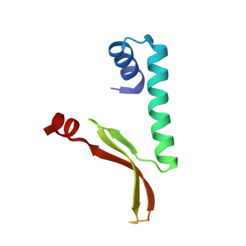
The Escherichia coli histone-like HU protein pool is composed of three dimeric forms: two homodimers, EcHUalpha(2) and EcHUbeta(2), and a heterodimer, EcHUalphabeta. The relative abundance of these dimeric forms varies during cell growth and in response to environmental changes, suggesting that each dimer plays different physiological roles. Here, differential scanning calorimetry and circular dichroism (CD) were used to study the thermal stability of the three E.coli HU dimers and show that each of them has its own thermodynamic signature. Unlike the other HU proteins studied so far, which melt through a single step (N(2)<-->2D), this present thermodynamic study shows that the three E.coli dimers melt according to a two-step mechanism (N(2)<-->I(2)<-->2D). The native dimer, N(2), melts partially into a dimeric intermediate, I(2), which in turn yields the unfolded monomers, D. In addition, the crystal structure of the EcHUalpha(2) dimer has been solved. Comparative thermodynamic and structural analysis between EcHUalpha(2) and the HU homodimer from Bacillus stearothermophilus suggests that the E.coli dimer is constituted by two subdomains of different energetic properties. The CD study indicates that the intermediate, I(2), corresponds to an HU dimer having partly lost its alpha-helices. The partially unfolded dimer I(2) is unable to complex with high-affinity, single-stranded break-containing DNA. These structural, thermodynamic and functional results suggest that the N(2)<-->I(2) equilibrium plays a central role in the physiology of E.coli HU. The I(2) molecular species seems to be the EcHUbeta(2) preferential conformation, possibly related to its role in the E.coli cold-shock adaptation. Besides, I(2) might be required in E.coli for the HU chain exchange, which allows the heterodimer formation from homodimers.
Organizational Affiliation:
Centre de Biophysique Moléculaire, CNRS, affiliated to the University of Orléans, rue Charles Sadron, 45071 Orléans cedex 02, France.