1MT3

Crystal Structure of the Tricorn Interacting Factor Selenomethionine-F1


Experimental Data Snapshot

  • Method: X-RAY DIFFRACTION
  • Resolution: 2.00 Å
  • R-Value Free: 0.280 
  • R-Value Work: 0.255 
  • R-Value Observed: 0.285 

wwPDB Validation   3D Report Full Report


This is version 1.2 of the entry. See complete history


Literature

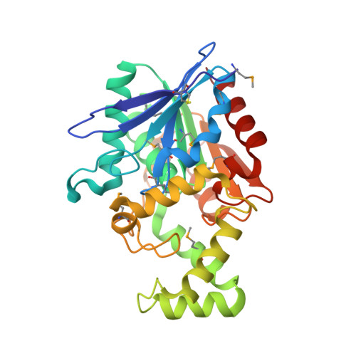
Structures of the tricorn-interacting aminopeptidase F1 with different ligands explain its catalytic mechanism

Goettig, P.Groll, M.Kim, J.-S.Huber, R.Brandstetter, H.

(2002) EMBO J 21: 5343-5352

  • DOI: https://doi.org/10.1093/emboj/cdf552
  • Primary Citation of Related Structures:  
    1MT3, 1MTZ, 1MU0

  • PubMed Abstract: 

    F1 is a 33.5 kDa serine peptidase of the alpha/beta-hydrolase family from the archaeon Thermoplasma acidophilum. Subsequent to proteasomal protein degradation, tricorn generates small peptides, which are cleaved by F1 to yield single amino acids. We have solved the crystal structure of F1 with multiwavelength anomalous dispersion (MAD) phasing at 1.8 A resolution. In addition to the conserved catalytic domain, the structure reveals a chiefly alpha-helical domain capping the catalytic triad. Thus, the active site is accessible only through a narrow opening from the protein surface. Two structures with molecules bound to the active serine, including the inhibitor phenylalanyl chloromethylketone, elucidate the N-terminal recognition of substrates and the catalytic activation switch mechanism of F1. The cap domain mainly confers the specificity for hydrophobic side chains by a novel cavity system, which, analogously to the tricorn protease, guides substrates to the buried active site and products away from it. Finally, the structure of F1 suggests a possible functional complex with tricorn that allows efficient processive degradation to free amino acids for cellular recycling.


  • Organizational Affiliation

    Max-Planck-Institut für Biochemie, Abteilung Strukturforschung, Am Klopferspitz 18a, D-82152 Martinsried, Germany. goettig@biochem.mpg.de


Macromolecules
Find similar proteins by:  (by identity cutoff)  |  3D Structure
Entity ID: 1
MoleculeChains Sequence LengthOrganismDetailsImage
Proline iminopeptidase293Thermoplasma acidophilumMutation(s): 9 
Gene Names: TA0830
EC: 3.4.11.5
UniProt
Find proteins for P96084 (Thermoplasma acidophilum (strain ATCC 25905 / DSM 1728 / JCM 9062 / NBRC 15155 / AMRC-C165))
Explore P96084 
Go to UniProtKB:  P96084
Entity Groups  
Sequence Clusters30% Identity50% Identity70% Identity90% Identity95% Identity100% Identity
UniProt GroupP96084
Sequence Annotations
Expand
  • Reference Sequence
Small Molecules
Ligands 1 Unique
IDChains Name / Formula / InChI Key2D Diagram3D Interactions
MES
Query on MES

Download Ideal Coordinates CCD File 
B [auth A]2-(N-MORPHOLINO)-ETHANESULFONIC ACID
C6 H13 N O4 S
SXGZJKUKBWWHRA-UHFFFAOYSA-N
Modified Residues  1 Unique
IDChains TypeFormula2D DiagramParent
MSE
Query on MSE
A
L-PEPTIDE LINKINGC5 H11 N O2 SeMET
Experimental Data & Validation

Experimental Data

  • Method: X-RAY DIFFRACTION
  • Resolution: 2.00 Å
  • R-Value Free: 0.280 
  • R-Value Work: 0.255 
  • R-Value Observed: 0.285 
  • Space Group: P 21 21 21
Unit Cell:
Length ( Å )Angle ( ˚ )
a = 51.968α = 90
b = 60.759β = 90
c = 80.678γ = 90
Software Package:
Software NamePurpose
DENZOdata reduction
SCALEPACKdata scaling
SHARPphasing
CNSrefinement

Structure Validation

View Full Validation Report


Entry History 

Deposition Data

Revision History  (Full details and data files)

  • Version 1.0: 2002-11-06
    Type: Initial release
  • Version 1.1: 2008-04-28
    Changes: Version format compliance
  • Version 1.2: 2011-07-13
    Changes: Version format compliance