1MR1

Crystal Structure of a Smad4-Ski Complex


Experimental Data Snapshot

  • Method: X-RAY DIFFRACTION
  • Resolution: 2.85 Å
  • R-Value Free: 0.280 
  • R-Value Work: 0.231 
  • R-Value Observed: 0.235 

wwPDB Validation   3D Report Full Report


This is version 1.3 of the entry. See complete history


Literature

Structural Mechanism of Smad4 Recognition by the Nuclear Oncoprotein Ski: Insights on Ski-mediated Repression of TGF-beta Signaling

Wu, J.-W.Krawitz, A.R.Chai, J.Li, W.Zhang, F.Luo, K.Shi, Y.

(2002) Cell 111: 357-367

  • DOI: https://doi.org/10.1016/s0092-8674(02)01006-1
  • Primary Citation of Related Structures:  
    1MR1

  • PubMed Abstract: 

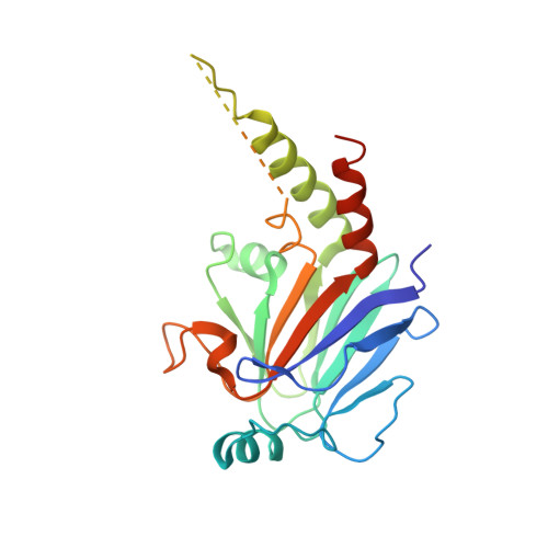
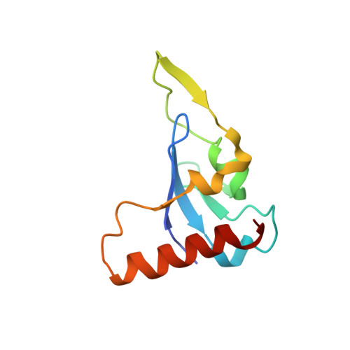
    The Ski family of nuclear oncoproteins represses TGF-beta signaling through interactions with the Smad proteins. The crystal structure of the Smad4 binding domain of human c-Ski in complex with the MH2 domain of Smad4 reveals specific recognition of the Smad4 L3 loop region by a highly conserved interaction loop (I loop) from Ski. The Ski binding surface on Smad4 significantly overlaps with that required for binding of the R-Smads. Indeed, Ski disrupts the formation of a functional complex between the Co- and R-Smads, explaining how it could lead to repression of TGF-beta, activin, and BMP responses. Intriguingly, the structure of the Ski fragment, stabilized by a bound zinc atom, resembles the SAND domain, in which the corresponding I loop is responsible for DNA binding.


  • Organizational Affiliation

    Department of Molecular Biology, Lewis Thomas Laboratory, Princeton University, Princeton, NJ 08544, USA.


Macromolecules
Find similar proteins by:  (by identity cutoff)  |  3D Structure
Entity ID: 1
MoleculeChains Sequence LengthOrganismDetailsImage
Mothers against decapentaplegic homolog 4
A, B
235Homo sapiensMutation(s): 0 
UniProt & NIH Common Fund Data Resources
Find proteins for Q13485 (Homo sapiens)
Explore Q13485 
Go to UniProtKB:  Q13485
PHAROS:  Q13485
GTEx:  ENSG00000141646 
Entity Groups  
Sequence Clusters30% Identity50% Identity70% Identity90% Identity95% Identity100% Identity
UniProt GroupQ13485
Sequence Annotations
Expand
  • Reference Sequence
Find similar proteins by:  (by identity cutoff)  |  3D Structure
Entity ID: 2
MoleculeChains Sequence LengthOrganismDetailsImage
Ski oncogene
C, D
99Homo sapiensMutation(s): 0 
UniProt & NIH Common Fund Data Resources
Find proteins for P12755 (Homo sapiens)
Explore P12755 
Go to UniProtKB:  P12755
PHAROS:  P12755
GTEx:  ENSG00000157933 
Entity Groups  
Sequence Clusters30% Identity50% Identity70% Identity90% Identity95% Identity100% Identity
UniProt GroupP12755
Sequence Annotations
Expand
  • Reference Sequence
Small Molecules
Ligands 1 Unique
IDChains Name / Formula / InChI Key2D Diagram3D Interactions
ZN
Query on ZN

Download Ideal Coordinates CCD File 
E [auth C],
F [auth D]
ZINC ION
Zn
PTFCDOFLOPIGGS-UHFFFAOYSA-N
Experimental Data & Validation

Experimental Data

  • Method: X-RAY DIFFRACTION
  • Resolution: 2.85 Å
  • R-Value Free: 0.280 
  • R-Value Work: 0.231 
  • R-Value Observed: 0.235 
  • Space Group: P 32 2 1
Unit Cell:
Length ( Å )Angle ( ˚ )
a = 109.8α = 90
b = 109.8β = 90
c = 141.1γ = 120
Software Package:
Software NamePurpose
DENZOdata reduction
SCALEPACKdata scaling
AMoREphasing
CNSrefinement

Structure Validation

View Full Validation Report


Entry History 

Deposition Data

Revision History  (Full details and data files)

  • Version 1.0: 2003-01-21
    Type: Initial release
  • Version 1.1: 2008-05-05
    Changes: Version format compliance
  • Version 1.2: 2011-07-13
    Changes: Version format compliance
  • Version 1.3: 2024-02-14
    Changes: Data collection, Database references, Derived calculations