The refined atomic structure of carbonic anhydrase II at 1.05 A resolution: implications of chemical rescue of proton transfer.
Duda, D., Govindasamy, L., Agbandje-McKenna, M., Tu, C., Silverman, D.N., McKenna, R.(2003) Acta Crystallogr D Biol Crystallogr 59: 93-104
- PubMed: 12499545
- DOI: https://doi.org/10.1107/s0907444902019455
- Primary Citation of Related Structures:
1MOO - PubMed Abstract:
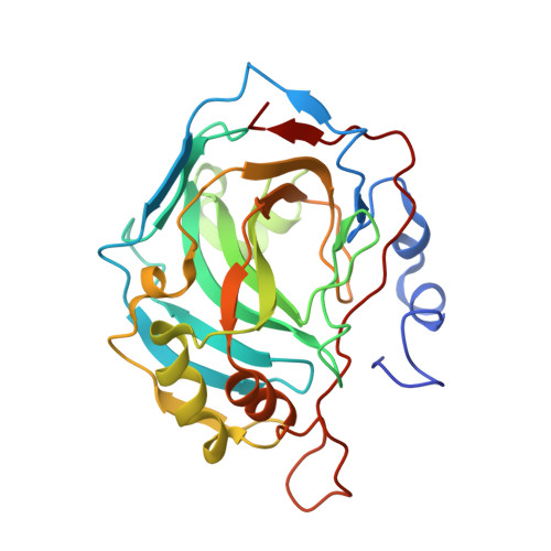
Using synchrotron radiation and a CCD detector, X-ray data have been collected at 100 K for the His64Ala mutant of human carbonic anhydrase II complexed with 4-methylimidazole (4-MI) to a maximal 1.05 A resolution, allowing full anisotropic least-squares refinement. The refined model has a conventional R factor of 15.7% for all reflections. The C(alpha) coordinates of the model presented here have an r.m.s. deviation of 0.10 A relative to the previously determined structure at 1.6 A resolution. Several amino-acid residues (six of the 255 observed) have been identified with multiple rotamer side-chain conformations. C, N and O atoms can be differentiated with selective electron-density map contouring. The estimated standard deviations for all main-chain non-H atom bond lengths and angles are 0.013 and 0.030 A, respectively, based on unrestrained full-matrix least-squares refinement. This structure gives detailed information about the tetrahedrally arranged zinc ion coordinated by three histidine N atoms (His94 N(epsilon 2), His96 N(epsilon2) and His119 N(delta1)) and a water/hydroxide, the multiple binding sites of the proton chemical rescue molecule 4-MI and the solvent networks linking the zinc-bound water/hydroxide and 4-MI molecules. This structure presents the highest resolution structure of a carbonic anhydrase isozyme so far determined and adds to the understanding of proton-transfer processes.
Organizational Affiliation:
Department of Biochemistry and Molecular Biology, University of Florida, Gainesville, FL 32610, USA.