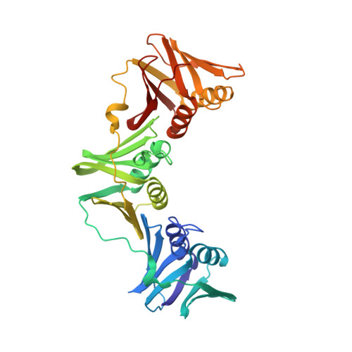
Flexibility revealed by the 1.85 A crystal structure of the beta sliding-clamp subunit of Escherichia coli DNA polymerase III.
Oakley, A.J., Prosselkov, P., Wijffels, G., Beck, J.L., Wilce, M.C., Dixon, N.E.(2003) Acta Crystallogr D Biol Crystallogr 59: 1192-1199
- PubMed: 12832762
- DOI: https://doi.org/10.1107/s0907444903009958
- Primary Citation of Related Structures:
1MMI - PubMed Abstract:
The beta subunit of the Escherichia coli replicative DNA polymerase III holoenzyme is the sliding clamp that interacts with the alpha (polymerase) subunit to maintain the high processivity of the enzyme. The beta protein is a ring-shaped dimer of 40.6 kDa subunits whose structure has previously been determined at a resolution of 2.5 A [Kong et al. (1992), Cell, 69, 425-437]. Here, the construction of a new plasmid that directs overproduction of beta to very high levels and a simple procedure for large-scale purification of the protein are described. Crystals grown under slightly modified conditions diffracted to beyond 1.9 A at 100 K at a synchrotron source. The structure of the beta dimer solved at 1.85 A resolution shows some differences from that reported previously. In particular, it was possible at this resolution to identify residues that differed in position between the two subunits in the unit cell; side chains of these and some other residues were found to occupy alternate conformations. This suggests that these residues are likely to be relatively mobile in solution. Some implications of this flexibility for the function of beta are discussed.
Organizational Affiliation:
Crystallography Centre, University of Western Australia, Nedlands, WA 6907, Australia.