Protein engineering with monomeric triosephosphate isomerase (monoTIM): the modelling and structure verification of a seven-residue loop.
Thanki, N., Zeelen, J.P., Mathieu, M., Jaenicke, R., Abagyan, R.A., Wierenga, R.K., Schliebs, W.(1997) Protein Eng 10: 159-167
- PubMed: 9089815
- DOI: https://doi.org/10.1093/protein/10.2.159
- Primary Citation of Related Structures:
1ML1 - PubMed Abstract:
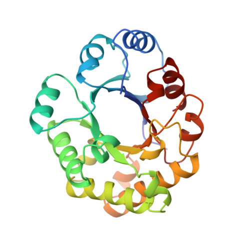
Protein engineering experiments have been carried out with loop-1 of monomeric triosephosphate isomerase (monoTIM). Loop-1 of monoTIM is disordered in every crystal structure of liganded monoTIM, but in the wild-type TIM it is a very rigid dimer interface loop. This loop connects the first beta-strand with the first alpha-helix of the TIM-barrel scaffold. The first residue of this loop, Lys13, is a conserved catalytic residue. The protein design studies with loop-1 were aimed at rigidifying this loop such that the Lys13 side chain points in the same direction as seen in wild type. The modelling suggested that the loop should be made one residue shorter. With the modelling package ICM the optimal sequence of a new seven-residue loop-1 was determined and its structure was predicted. The new variant could be expressed and purified and has been characterized. The catalytic activity and stability are very similar to those of monoTIM. The crystal structure (at 2.6 A resolution) shows that the experimental loop-1 structure agrees well with the modelled loop-1 structure. The direct superposition of the seven loop residues of the modelled and experimental structures results in an r.m.s. difference of 0.5 A for the 28 main chain atoms. The good agreement between the predicted structure and the crystal structure shows that the described modelling protocol can be used successfully for the reliable prediction of loop structures.
Organizational Affiliation:
Institut für Biophysik und Physikalische Biochemie, Universitt Regensburg, Germany.