1MKD

crystal structure of PDE4D catalytic domain and zardaverine complex


Experimental Data Snapshot

  • Method: X-RAY DIFFRACTION
  • Resolution: 2.90 Å
  • R-Value Free: 0.260 
  • R-Value Work: 0.245 
  • R-Value Observed: 0.245 

wwPDB Validation   3D Report Full Report


This is version 1.3 of the entry. See complete history


Literature

Crystal structure of phosphodiesterase 4D and inhibitor complex

Lee, M.E.Markowitz, J.Lee, J.-O.Lee, H.

(2002) FEBS Lett 530: 53-58

  • DOI: https://doi.org/10.1016/s0014-5793(02)03396-3
  • Primary Citation of Related Structures:  
    1MKD

  • PubMed Abstract: 

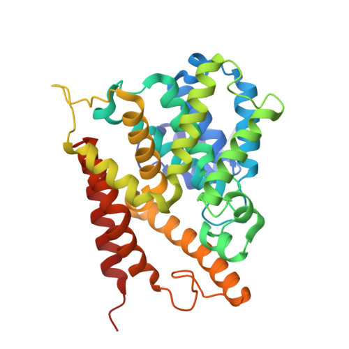
    Cyclic nucleotide phosphodiesterases (PDEs) regulate physiological processes by degrading intracellular second messengers, adenosine-3',5'-cyclic phosphate or guanosine-3',5'-cyclic phosphate. The first crystal structure of PDE4D catalytic domain and a bound inhibitor, zardaverine, was determined. Zardaverine binds to a highly conserved pocket that includes the catalytic metal binding site. Zardaverine fills only a portion of the active site pocket. More selective PDE4 inhibitors including rolipram, cilomilast and roflumilast have additional functional groups that can utilize the remaining empty space for increased binding energy and selectivity. In the crystal structure, the catalytic domain of PDE4D possesses an extensive dimerization interface containing residues that are highly conserved in PDE1, 3, 4, 8 and 9. Mutations of R358D or D322R among these interface residues prohibit dimerization of the PDE4D catalytic domain in solution.


  • Organizational Affiliation

    Department of Chemistry, Korea Advanced Institute of Science and Technology, 373-1 Kusong-dong, Yusong-gu, Daejon 305-701, South Korea.


Macromolecules
Find similar proteins by:  (by identity cutoff)  |  3D Structure
Entity ID: 1
MoleculeChains Sequence LengthOrganismDetailsImage
Phosphodiesterase 4D
A, B, C, D, E
A, B, C, D, E, F, G, H, I, J, K, L
328Homo sapiensMutation(s): 0 
EC: 3.1.4.17
UniProt & NIH Common Fund Data Resources
Find proteins for Q08499 (Homo sapiens)
Explore Q08499 
Go to UniProtKB:  Q08499
PHAROS:  Q08499
GTEx:  ENSG00000113448 
Entity Groups  
Sequence Clusters30% Identity50% Identity70% Identity90% Identity95% Identity100% Identity
UniProt GroupQ08499
Sequence Annotations
Expand
  • Reference Sequence
Small Molecules
Ligands 3 Unique
IDChains Name / Formula / InChI Key2D Diagram3D Interactions
ZAR
Query on ZAR

Download Ideal Coordinates CCD File 
AA [auth E]
DA [auth F]
GA [auth G]
JA [auth H]
MA [auth I]
AA [auth E],
DA [auth F],
GA [auth G],
JA [auth H],
MA [auth I],
O [auth A],
PA [auth J],
R [auth B],
SA [auth K],
U [auth C],
VA [auth L],
X [auth D]
6-(4-DIFLUOROMETHOXY-3-METHOXY-PHENYL)-2H-PYRIDAZIN-3-ONE
C12 H10 F2 N2 O3
HJMQDJPMQIHLPB-UHFFFAOYSA-N
ZN
Query on ZN

Download Ideal Coordinates CCD File 
BA [auth F]
EA [auth G]
HA [auth H]
KA [auth I]
M [auth A]
BA [auth F],
EA [auth G],
HA [auth H],
KA [auth I],
M [auth A],
NA [auth J],
P [auth B],
QA [auth K],
S [auth C],
TA [auth L],
V [auth D],
Y [auth E]
ZINC ION
Zn
PTFCDOFLOPIGGS-UHFFFAOYSA-N
MG
Query on MG

Download Ideal Coordinates CCD File 
CA [auth F]
FA [auth G]
IA [auth H]
LA [auth I]
N [auth A]
CA [auth F],
FA [auth G],
IA [auth H],
LA [auth I],
N [auth A],
OA [auth J],
Q [auth B],
RA [auth K],
T [auth C],
UA [auth L],
W [auth D],
Z [auth E]
MAGNESIUM ION
Mg
JLVVSXFLKOJNIY-UHFFFAOYSA-N
Binding Affinity Annotations 
IDSourceBinding Affinity
ZAR BindingDB:  1MKD IC50: min: 158, max: 2000 (nM) from 9 assay(s)
PDBBind:  1MKD IC50: 160 (nM) from 1 assay(s)
Experimental Data & Validation

Experimental Data

  • Method: X-RAY DIFFRACTION
  • Resolution: 2.90 Å
  • R-Value Free: 0.260 
  • R-Value Work: 0.245 
  • R-Value Observed: 0.245 
  • Space Group: P 21 21 21
Unit Cell:
Length ( Å )Angle ( ˚ )
a = 97.325α = 90
b = 164.572β = 90
c = 325.54γ = 90
Software Package:
Software NamePurpose
DENZOdata reduction
SCALEPACKdata scaling
AMoREphasing
CNSrefinement

Structure Validation

View Full Validation Report


Entry History 

Deposition Data

Revision History  (Full details and data files)

  • Version 1.0: 2003-03-01
    Type: Initial release
  • Version 1.1: 2008-04-28
    Changes: Version format compliance
  • Version 1.2: 2011-07-13
    Changes: Version format compliance
  • Version 1.3: 2024-03-13
    Changes: Data collection, Database references, Derived calculations