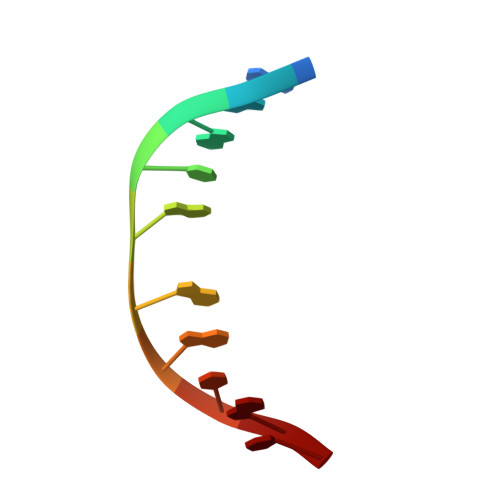
1MK6

SOLUTION STRUCTURE OF THE 8,9-DIHYDRO-8-(N7-GUANYL)-9-HYDROXY-AFLATOXIN B1 ADDUCT MISPAIRED WITH DEOXYADENOSINE


Experimental Data Snapshot

  • Method: SOLUTION NMR
  • Conformers Calculated: 20 
  • Conformers Submitted: 
  • Selection Criteria: Final Calculated Structure is Being Submitted. Back Calculated Structure is in Agreement with NOESY data. The calculation was performed in Presence of solvent and counterions. Solvent, and Counterion co-ordinates are NOT being reported, only the Duplex DNA. Before Solvating and Addition of Counter IONS, 20 final structures were calculated using XPLOR. The final averaged energy minimized structure was solvated, and the counter Ions were added to it. Then MD was ran for 1.4 ns time scale to obtain final structure. 

wwPDB Validation   3D Report Full Report


This is version 2.0 of the entry. See complete history


Literature

MISPAIRING OF THE 8,9-DIHYDRO-8-(N7-GUANYL)-9-HYDROXY-AFLATOXIN B1 ADDUCT WITH DEOXYADENOSINE RESULTS IN EXTRUSION OF THE MISMATCHED DA TOWARD THE MAJOR GROOVE

Giri, I.Johnston, D.S.Stone, M.P.

(2002) Biochemistry 41: 5462-5472

  • DOI: https://doi.org/10.1021/bi012116t
  • Primary Citation of Related Structures:  
    1MK6

  • PubMed Abstract: 

    The G --> T transversion is the dominant mutation induced by the cationic trans-8,9-dihydro-8-(N7-guanyl)-9-hydroxy-aflatoxin B(1) adduct. The structure of d(ACATC(AFB)GATCT).d(AGATAGATGT), in which the cationic adduct was mismatched with deoxyadenosine, was refined using molecular dynamics calculations restrained by NOE data and dihedral restraints obtained from NMR spectroscopy. Restrained molecular dynamics calculations refined structures with pairwise rmsd <1 A and a sixth root R1x factor between the refined structure and NOE data of 10.5 x 10⁻². The mismatched duplex existed in a single conformation at neutral pH. The aflatoxin moiety intercalated above the 5' face of the modified (AFB)G. The mismatched dA was in the anti conformation about the glycosyl bond. It extruded toward the major groove and did not participate in hydrogen bonding with (AFB)G. The structure was compared with that of d(ACATCGATCT).d(AGATAGATGT) containing the corresponding unmodified G.A mismatch and with d(ACATC(AFB)GATCT).d(AGATCGATGT) containing the aflatoxin lesion in the correctly paired (AFB)G.C context. The correctly paired oligodeoxynucleotide exhibited Watson-Crick-type geometry at the (AFB)G.C pair. It melted at higher temperature than the mismatched (AFB)G.A duplex. The unmodified mismatched G.A duplex exhibited spectral line broadening at neutral pH, suggesting a mixture of conformations. It exhibited a lower melting temperature than did the mismatched (AFB)G.A duplex. These differences correlated with replication bypass experiments performed in vitro utilizing DNA polymerase I exo- [Johnston, D. S., and Stone, M. P. (2000) Chem. Res. Toxicol. 13, 1158-1164]. Those experiments showed that correct insertion of dC opposite (AFB)G blocked replication by the enzyme, whereas incorrect insertion of dA opposite (AFB)G allowed full-length replication of the adducted template strand.


  • Organizational Affiliation

    Department of Chemistry and Center in Molecular Toxicology, Vanderbilt University, Nashville, Tennessee 37235, USA.


Macromolecules

Find similar nucleic acids by:  Sequence   |   3D Structure  

Entity ID: 1
MoleculeChains LengthOrganismImage
5'-D(*AP*CP*AP*TP*CP*GP*AP*TP*CP*T)-3'10synthetic construct
Sequence Annotations
Expand
  • Reference Sequence

Find similar nucleic acids by:  Sequence   |   3D Structure  

Entity ID: 2
MoleculeChains LengthOrganismImage
5'-D(*AP*GP*AP*TP*AP*GP*AP*TP*GP*T)-3'10synthetic construct
Sequence Annotations
Expand
  • Reference Sequence
Small Molecules
Ligands 1 Unique
IDChains Name / Formula / InChI Key2D Diagram3D Interactions
AFN
Query on AFN

Download Ideal Coordinates CCD File 
C [auth A]8,9-DIHYDRO-9-HYDROXY-AFLATOXIN B1
C17 H14 O7
GACWFTLPANAFAK-GVLSTKRJSA-N
Experimental Data & Validation

Experimental Data

  • Method: SOLUTION NMR
  • Conformers Calculated: 20 
  • Conformers Submitted: 
  • Selection Criteria: Final Calculated Structure is Being Submitted. Back Calculated Structure is in Agreement with NOESY data. The calculation was performed in Presence of solvent and counterions. Solvent, and Counterion co-ordinates are NOT being reported, only the Duplex DNA. Before Solvating and Addition of Counter IONS, 20 final structures were calculated using XPLOR. The final averaged energy minimized structure was solvated, and the counter Ions were added to it. Then MD was ran for 1.4 ns time scale to obtain final structure. 

Structure Validation

View Full Validation Report


Entry History 

Deposition Data

Revision History  (Full details and data files)

  • Version 1.0: 2002-10-16
    Type: Initial release
  • Version 1.1: 2008-04-28
    Changes: Version format compliance
  • Version 1.2: 2011-07-13
    Changes: Version format compliance
  • Version 2.0: 2019-01-30
    Changes: Atomic model, Data collection, Database references, Derived calculations, Non-polymer description, Polymer sequence, Source and taxonomy, Structure summary