1MJX

STRUCTURE OF INORGANIC PYROPHOSPHATASE MUTANT D65N


Experimental Data Snapshot

  • Method: X-RAY DIFFRACTION
  • Resolution: 2.15 Å
  • R-Value Free: 0.263 
  • R-Value Work: 0.190 

wwPDB Validation   3D Report Full Report


This is version 1.4 of the entry. See complete history


Literature

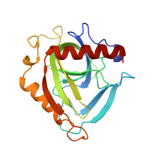
Three-dimensional structures of mutant forms of E. coli inorganic pyrophosphatase with Asp-->Asn single substitution in positions 42, 65, 70, and 97.

Avaeva, S.M.Rodina, E.V.Vorobyeva, N.N.Kurilova, S.A.Nazarova, T.I.Sklyankina, V.A.Oganessyan, V.Y.Samygina, V.R.Harutyunyan, E.H.

(1998) Biochemistry (Mosc) 63: 671-684

  • Primary Citation of Related Structures:  
    1MJW, 1MJX, 1MJY, 1MJZ

  • PubMed Abstract: 

    The three-dimensional structures of four mutant E. coli inorganic pyrophosphatases (PPases) with single Asp-->Asn substitutions at positions 42, 65, 70, and 97 were solved at 1.95, 2.15, 2.10, and 2.20 A resolution, respectively. Asp-42-->Asn and Asp-65-->Asn mutant PPases were prepared as complexes with sulfate--a structural analog of phosphate, the product of enzymatic reaction. A comparison of mutant enzymes with native PPases revealed that a single amino acid substitution changes the position of the mutated residue as well as the positions of several functional groups and some parts of a polypeptide chain. These changes are responsible for the fact that mutant PPases differ from the native ones in their catalytic properties. The sulfate binding to the mutant PPase active site causes molecular asymmetry, as shown for the native PPase earlier. The subunit asymmetry is manifested in different positions of sulfate and several functional groups, as well as changes in packing of hexamers in crystals and in cell parameters.


  • Organizational Affiliation

    Belozersky Institute of Physico-Chemical Biology, Lomonosov Moscow State University, Moscow, 119899, Russia. avaeva@protchem.genebee. msu.su.


Macromolecules
Find similar proteins by:  (by identity cutoff)  |  3D Structure
Entity ID: 1
MoleculeChains Sequence LengthOrganismDetailsImage
INORGANIC PYROPHOSPHATASE
A, B
175Escherichia coliMutation(s): 1 
EC: 3.6.1.1
UniProt
Find proteins for P0A7A9 (Escherichia coli (strain K12))
Explore P0A7A9 
Go to UniProtKB:  P0A7A9
Entity Groups  
Sequence Clusters30% Identity50% Identity70% Identity90% Identity95% Identity100% Identity
UniProt GroupP0A7A9
Sequence Annotations
Expand
  • Reference Sequence
Small Molecules
Ligands 1 Unique
IDChains Name / Formula / InChI Key2D Diagram3D Interactions
SO4
Query on SO4

Download Ideal Coordinates CCD File 
C [auth A],
D [auth B]
SULFATE ION
O4 S
QAOWNCQODCNURD-UHFFFAOYSA-L
Experimental Data & Validation

Experimental Data

  • Method: X-RAY DIFFRACTION
  • Resolution: 2.15 Å
  • R-Value Free: 0.263 
  • R-Value Work: 0.190 
  • Space Group: H 3 2
Unit Cell:
Length ( Å )Angle ( ˚ )
a = 110.2α = 90
b = 110.2β = 90
c = 155.2γ = 120
Software Package:
Software NamePurpose
REFMACrefinement

Structure Validation

View Full Validation Report


Entry History 

Deposition Data

Revision History  (Full details and data files)

  • Version 1.0: 1997-12-03
    Type: Initial release
  • Version 1.1: 2008-03-24
    Changes: Version format compliance
  • Version 1.2: 2011-07-13
    Changes: Derived calculations, Version format compliance
  • Version 1.3: 2021-11-03
    Changes: Database references, Derived calculations, Other
  • Version 1.4: 2024-02-14
    Changes: Data collection