1MJT

CRYSTAL STRUCTURE OF SANOS, A BACTERIAL NITRIC OXIDE SYNTHASE OXYGENASE PROTEIN, IN COMPLEX WITH NAD+ AND SEITU


Experimental Data Snapshot

  • Method: X-RAY DIFFRACTION
  • Resolution: 2.40 Å
  • R-Value Free: 0.241 
  • R-Value Work: 0.182 
  • R-Value Observed: 0.177 

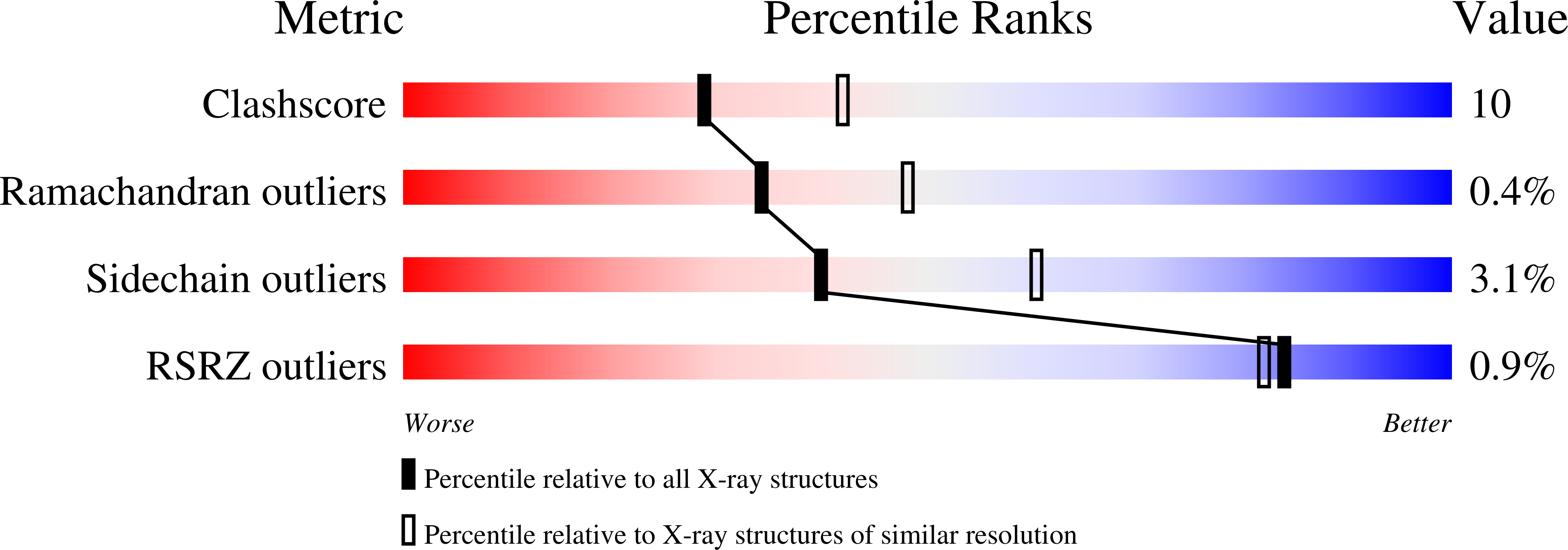
wwPDB Validation   3D Report Full Report


Ligand Structure Quality Assessment 


This is version 2.1 of the entry. See complete history


Literature

Crystal Structure of SANOS, a Bacterial Nitric Oxide Synthase Oxygenase Protein from Staphylococcus aureus

Bird, L.E.Ren, J.Zhang, J.Foxwell, N.Hawkins, A.R.Charles, I.G.Stammers, D.K.

(2002) Structure 10: 1687-1696

  • DOI: https://doi.org/10.1016/s0969-2126(02)00911-5
  • Primary Citation of Related Structures:  
    1MJT

  • PubMed Abstract: 

    Prokaryotic genes related to the oxygenase domain of mammalian nitric oxide synthases (NOSs) have recently been identified. Although they catalyze the same reaction as the eukaryotic NOS oxygenase domain, their biological function(s) are unknown. In order to explore rationally the biochemistry and evolution of the prokaryotic NOS family, we have determined the crystal structure of SANOS, from methicillin-resistant Staphylococcus aureus (MRSA), to 2.4 A. Haem and S-ethylisothiourea (SEITU) are bound at the SANOS active site, while the intersubunit site, occupied by the redox cofactor tetrahydrobiopterin (H(4)B) in mammalian NOSs, has NAD(+) bound in SANOS. In common with all bacterial NOSs, SANOS lacks the N-terminal extension responsible for stable dimerization in mammalian isoforms, but has alternative interactions to promote dimer formation.


  • Organizational Affiliation

    Division of Structural Biology, The Wellcome Trust Centre for Human Genetics, Henry Wellcome Building of Genomic Medicine, University of Oxford, Roosevelt Drive, Oxford OX3 7BN, United Kingdom.


Macromolecules
Find similar proteins by:  (by identity cutoff)  |  3D Structure
Entity ID: 1
MoleculeChains Sequence LengthOrganismDetailsImage
NITRIC-OXIDE SYNTHASE HOMOLOG
A, B
347Staphylococcus aureusMutation(s): 0 
Gene Names: sanos
EC: 1.14.13.39
UniProt
Find proteins for P0A004 (Staphylococcus aureus)
Explore P0A004 
Go to UniProtKB:  P0A004
Entity Groups  
Sequence Clusters30% Identity50% Identity70% Identity90% Identity95% Identity100% Identity
UniProt GroupP0A004
Sequence Annotations
Expand
  • Reference Sequence
Oligosaccharides

Help

Entity ID: 2
MoleculeChains Length2D Diagram Glycosylation3D Interactions
beta-D-fructofuranose-(2-1)-alpha-D-glucopyranose
C, D
2N/A
Glycosylation Resources
GlyTouCan:  G05551OP
GlyCosmos:  G05551OP
Biologically Interesting Molecules (External Reference) 1 Unique
Experimental Data & Validation

Experimental Data

  • Method: X-RAY DIFFRACTION
  • Resolution: 2.40 Å
  • R-Value Free: 0.241 
  • R-Value Work: 0.182 
  • R-Value Observed: 0.177 
  • Space Group: P 21 21 21
Unit Cell:
Length ( Å )Angle ( ˚ )
a = 65.78α = 90
b = 115.14β = 90
c = 126.02γ = 90
Software Package:
Software NamePurpose
DENZOdata reduction
SCALEPACKdata scaling
CNSrefinement
CNSphasing

Structure Validation

View Full Validation Report


Ligand Structure Quality Assessment 


Entry History 

Deposition Data

Revision History  (Full details and data files)

  • Version 1.0: 2003-01-07
    Type: Initial release
  • Version 1.1: 2008-04-28
    Changes: Version format compliance
  • Version 1.2: 2011-07-13
    Changes: Version format compliance
  • Version 2.0: 2020-07-29
    Type: Remediation
    Reason: Carbohydrate remediation
    Changes: Atomic model, Data collection, Database references, Derived calculations, Non-polymer description, Structure summary
  • Version 2.1: 2024-02-14
    Changes: Data collection, Database references, Refinement description, Structure summary