1MJC

CRYSTAL STRUCTURE OF CSPA, THE MAJOR COLD SHOCK PROTEIN OF ESCHERICHIA COLI


Experimental Data Snapshot

  • Method: X-RAY DIFFRACTION
  • Resolution: 2.00 Å
  • R-Value Work: 0.187 
  • R-Value Observed: 0.187 

wwPDB Validation   3D Report Full Report


This is version 1.6 of the entry. See complete history


Literature

Crystal structure of CspA, the major cold shock protein of Escherichia coli.

Schindelin, H.Jiang, W.Inouye, M.Heinemann, U.

(1994) Proc Natl Acad Sci U S A 91: 5119-5123

  • DOI: https://doi.org/10.1073/pnas.91.11.5119
  • Primary Citation of Related Structures:  
    1MJC

  • PubMed Abstract: 

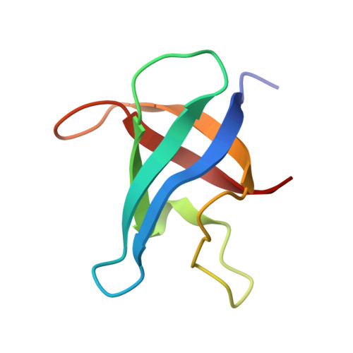
    The major cold shock protein of Escherichia coli, CspA, produced upon a rapid downshift in growth temperature, is involved in the transcriptional regulation of at least two genes. The protein shares high homology with the nucleic acid-binding domain of the Y-box factors, a family of eukaryotic proteins involved in transcriptional and translational regulation. The crystal structure of CspA has been determined at 2-A resolution and refined to R = 0.187. CspA is composed of five antiparallel beta-strands forming a closed five-stranded beta-barrel. The three-dimensional structure of CspA is similar to that of the major cold shock protein of Bacillus subtilis, CspB, which has recently been determined at 2.45-A resolution. However, in contrast to CspB, no dimer is formed in the crystal. The surface of CspA is characteristic for a protein interacting with single-stranded nucleic acids. Due to the high homology of the bacterial cold shock proteins with the Y-box factors, E. coli CspA and B. subtilis CspB define a structural framework for the common cold shock domain.


  • Organizational Affiliation

    Forschungsgruppe Kristallographie, Max-Delbrúck-Centrum für Molekulare Medizin, Berlin, Germany.


Macromolecules
Find similar proteins by:  (by identity cutoff)  |  3D Structure
Entity ID: 1
MoleculeChains Sequence LengthOrganismDetailsImage
MAJOR COLD-SHOCK PROTEIN 7.469Escherichia coliMutation(s): 0 
UniProt
Find proteins for P0A9X9 (Escherichia coli (strain K12))
Explore P0A9X9 
Go to UniProtKB:  P0A9X9
Entity Groups  
Sequence Clusters30% Identity50% Identity70% Identity90% Identity95% Identity100% Identity
UniProt GroupP0A9X9
Sequence Annotations
Expand
  • Reference Sequence
Experimental Data & Validation

Experimental Data

  • Method: X-RAY DIFFRACTION
  • Resolution: 2.00 Å
  • R-Value Work: 0.187 
  • R-Value Observed: 0.187 
  • Space Group: P 21 21 21
Unit Cell:
Length ( Å )Angle ( ˚ )
a = 47.12α = 90
b = 39.92β = 90
c = 30.92γ = 90
Software Package:
Software NamePurpose
X-PLORmodel building
PROLSQrefinement
X-PLORrefinement
X-PLORphasing

Structure Validation

View Full Validation Report


Entry History 

Deposition Data

Revision History  (Full details and data files)

  • Version 1.0: 1994-06-22
    Type: Initial release
  • Version 1.1: 2008-03-24
    Changes: Version format compliance
  • Version 1.2: 2011-07-13
    Changes: Version format compliance
  • Version 1.3: 2017-11-29
    Changes: Derived calculations, Other
  • Version 1.4: 2019-07-17
    Changes: Data collection, Refinement description
  • Version 1.5: 2019-08-14
    Changes: Data collection, Refinement description
  • Version 1.6: 2024-02-14
    Changes: Data collection, Database references