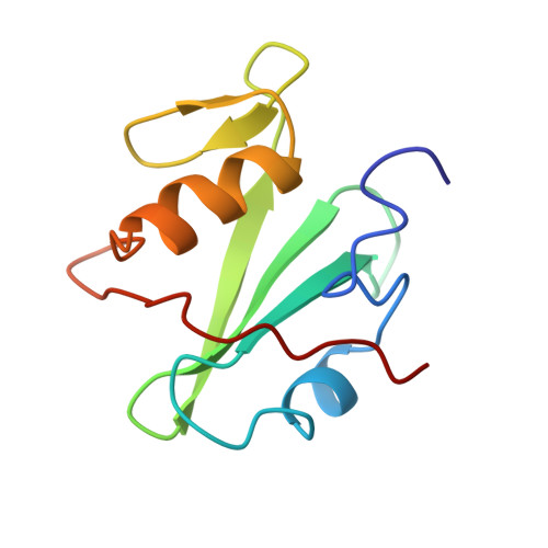
Crystal structure of the SH2 domain from the adaptor protein SHC: a model for peptide binding based on X-ray and NMR data.
Mikol, V., Baumann, G., Zurini, M.G., Hommel, U.(1995) J Mol Biol 254: 86-95
- PubMed: 7473762
- DOI: https://doi.org/10.1006/jmbi.1995.0601
- Primary Citation of Related Structures:
1MIL - PubMed Abstract:
Src homology 2 domains (SH2) are protein molecules found within a wide variety of cytoplasmic signalling molecules that bind with high affinity to phosphotyrosyl (pY)-containing protein sequences. We report here for crystal structure of the SH2 domain from the adaptor protein SHC (Shc), which has been refined by restrained least-squares methods to an R-factor of 17.3% to 2.7 A. The overall Shc architecture is essentially similar to that determined in other SH2 domains but it shows significant differences in a number of loops, thus providing a molecular surface with no obvious secondary pocket. Based on the knowledge of the crystal structure of the protein a model for a low affinity Shc-bound peptide has been generated from nuclear magnetic resonance data in solution using transferred nuclear Overhauser enhancements as intramolecular distance restraints. The model shows that the tyrosine moiety binds Shc in a rather similar way to that observed for other SH2-peptide complexes, but that the residue in position +3 does not seem to make specific contact with the protein. An intermolecular crystallographic interaction occurs between the pY-binding site and the C-terminal residues of a symmetry-related molecule. This crystal packing interaction suggests how inhibitory regulation could play a role in SHC activity.
Organizational Affiliation:
Preclinical Research, Sandoz Pharma AG, Basel Switzerland.