1MGO

Horse Liver Alcohol Dehydrogenase Phe93Ala Mutant


Experimental Data Snapshot

  • Method: X-RAY DIFFRACTION
  • Resolution: 1.20 Å
  • R-Value Free: 0.209 
  • R-Value Work: 0.188 
  • R-Value Observed: 0.188 

wwPDB Validation   3D Report Full Report


Ligand Structure Quality Assessment 


This is version 1.4 of the entry. See complete history


Literature

Mobility of Fluorobenzyl Alcohols Bound to Liver Alcohol Dehydrogenases as Determined by NMR and X-ray Crystallographic Studies

Rubach, J.K.Plapp, B.V.

(2002) Biochemistry 41: 15770-15779

  • DOI: https://doi.org/10.1021/bi026581h
  • Primary Citation of Related Structures:  
    1MG0, 1MGO

  • PubMed Abstract: 

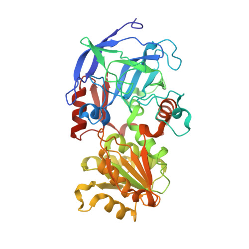
    The relationship between substrate mobility and catalysis was studied with wild-type and Phe93Ala (F93A) horse liver alcohol dehydrogenase (ADH). Wild-type ADH binds 2,3,4,5,6-pentafluorobenzyl alcohol in one position as shown by X-ray results, and (19)F NMR shows five resonances for the fluorines of the bound alcohol. The two meta-fluorines exchange positions with a rate constant of about 4 s(-1), indicating that mobility (ring flipping) of the benzyl alcohol is relatively restricted. The wild-type enzyme binds 2,3-difluorobenzyl alcohol in two alternative conformations that are related by a ring flip and a small translation of the fluorinated benzene ring, and the (19)F NMR spectrum shows three resonances for the two bound fluorines, consistent with the two orientations. Phe-93 interacts with the bound benzyl alcohols, and the F93A substitution decreases the rate constants for hydride transfer for benzyl alcohol oxidation and benzaldehyde reduction by 7.4- and 130-fold, respectively. The structure of F93A ADH crystallized with NAD(+) and 2,3,4,5,6-pentafluorobenzyl alcohol is similar to the structure of the wild-type enzyme complex except that the pentafluorobenzyl alcohol is not found in one position. The (19)F NMR spectrum of the F93A ADH-NAD(+)-pentafluorobenzyl alcohol complex shows three resonances for the bound fluorines. Line shape analysis of the spectrum suggests the bound pentafluorobenzyl ring undergoes rapid ring-flipping at about 20 000 s(-1). The F93A substitution greatly increases the mobility of the benzyl alcohol but modestly and differentially decreases the probability that the substrate is preorganized for hydride transfer.


  • Organizational Affiliation

    Department of Biochemistry, The University of Iowa, Iowa City, IA 52242, USA.


Macromolecules
Find similar proteins by:  (by identity cutoff)  |  3D Structure
Entity ID: 1
MoleculeChains Sequence LengthOrganismDetailsImage
Alcohol Dehydrogenase E chain
A, B
374Equus caballusMutation(s): 1 
Gene Names: M64864
EC: 1.1.1.1
UniProt
Find proteins for P00327 (Equus caballus)
Explore P00327 
Go to UniProtKB:  P00327
Entity Groups  
Sequence Clusters30% Identity50% Identity70% Identity90% Identity95% Identity100% Identity
UniProt GroupP00327
Sequence Annotations
Expand
  • Reference Sequence
Small Molecules
Ligands 4 Unique
IDChains Name / Formula / InChI Key2D Diagram3D Interactions
NAD
Query on NAD

Download Ideal Coordinates CCD File 
E [auth A],
H [auth B]
NICOTINAMIDE-ADENINE-DINUCLEOTIDE
C21 H27 N7 O14 P2
BAWFJGJZGIEFAR-NNYOXOHSSA-N
PFB
Query on PFB

Download Ideal Coordinates CCD File 
I [auth B],
J [auth B]
2,3,4,5,6-PENTAFLUOROBENZYL ALCOHOL
C7 H3 F5 O
PGJYYCIOYBZTPU-UHFFFAOYSA-N
MPD
Query on MPD

Download Ideal Coordinates CCD File 
K [auth B](4S)-2-METHYL-2,4-PENTANEDIOL
C6 H14 O2
SVTBMSDMJJWYQN-YFKPBYRVSA-N
ZN
Query on ZN

Download Ideal Coordinates CCD File 
C [auth A],
D [auth A],
F [auth B],
G [auth B]
ZINC ION
Zn
PTFCDOFLOPIGGS-UHFFFAOYSA-N
Binding Affinity Annotations 
IDSourceBinding Affinity
PFB Binding MOAD:  1MGO Ki: 5.10e+4 (nM) from 1 assay(s)
Experimental Data & Validation

Experimental Data

  • Method: X-RAY DIFFRACTION
  • Resolution: 1.20 Å
  • R-Value Free: 0.209 
  • R-Value Work: 0.188 
  • R-Value Observed: 0.188 
  • Space Group: P 1
Unit Cell:
Length ( Å )Angle ( ˚ )
a = 44.144α = 91.98
b = 51.308β = 103.01
c = 93.481γ = 109.81
Software Package:
Software NamePurpose
d*TREKdata scaling
d*TREKdata reduction
AMoREphasing
REFMACrefinement

Structure Validation

View Full Validation Report


Ligand Structure Quality Assessment 


Entry History 

Deposition Data

Revision History  (Full details and data files)

  • Version 1.0: 2002-11-13
    Type: Initial release
  • Version 1.1: 2008-04-28
    Changes: Version format compliance
  • Version 1.2: 2011-07-13
    Changes: Version format compliance
  • Version 1.3: 2021-10-27
    Changes: Database references, Derived calculations
  • Version 1.4: 2024-02-14
    Changes: Data collection, Refinement description