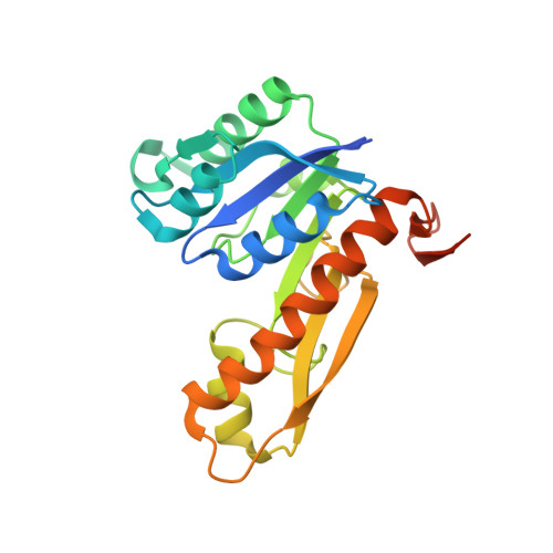
Crystal structures of human GAR Tfase of low and high pH and with substrate beta-GAR
Zhang, Y., Desharnais, J., Greasley, S.E., Beardsley, G.P., Boger, D.L., Wilson, I.A.(2002) Biochemistry 41: 14206-14215
- PubMed: 12450384
- DOI: https://doi.org/10.1021/bi020522m
- Primary Citation of Related Structures:
1MEJ, 1MEN, 1MEO - PubMed Abstract:
Glycinamide ribonucleotide transformylase (GAR Tfase) is a key folate-dependent enzyme in the de novo purine biosynthesis pathway and, as such, has been the target for antitumor drug design. Here, we describe the crystal structures of the human GAR Tfase (purN) component of the human trifunctional protein (purD-purM-purN) at various pH values and in complex with its substrate. Human GAR Tfase exhibits pH-dependent enzyme activity with its maximum around pH 7.5-8. Comparison of unliganded human GAR Tfase structures at pH 4.2 and pH 8.5 reveals conformational differences in the substrate binding loop, which at pH 4.2 occupies the binding cleft and prohibits substrate binding, while at pH 8.5 is permissive for substrate binding. The crystal structure of GAR Tfase with its natural substrate, beta-glycinamide ribonucleotide (beta-GAR), at pH 8.5 confirms this conformational isomerism. Surprisingly, several important structural differences are found between human GAR Tfase and previously reported E. coli GAR Tfase structures, which have been used as the primary template for drug design studies. While the E. coli structure gave valuable insights into the active site and formyl transfer mechanism, differences in structure and inhibition between the bacterial and mammalian enzymes suggest that the human GAR Tfase structure is now the appropriate template for the design of anti-cancer agents.
Organizational Affiliation:
Department of Molecular Biology, The Skaggs Institute for Chemical Biology, The Scripps Research Institute, 10550 North Torrey Pines Road, La Jolla, California 92037, USA.