A Calcium-driven Conformational Switch of the N-terminaland Core Domains of Annexin A1
Luecke, H., Rosengarth, A.(2003) J Mol Biol 326: 1317-1325
- PubMed: 12595246
- DOI: https://doi.org/10.1016/s0022-2836(03)00027-5
- Primary Citation of Related Structures:
1MCX - PubMed Abstract:
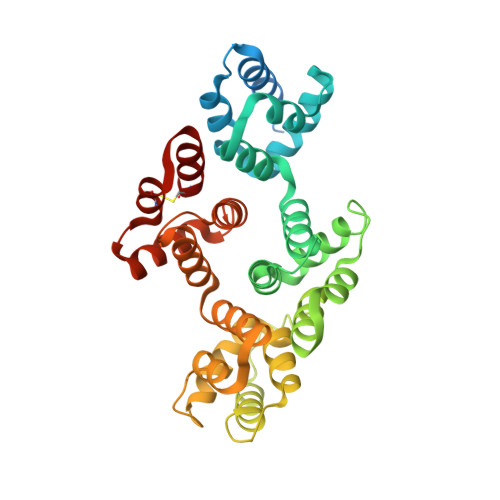
In 1993, Huber and co-workers published the structure of an N-terminally truncated version of human annexin A1 lacking the first 32 amino acid residues (PDB code: 1AIN). In 2001, we reported the structure of full-length porcine annexin A1 including the N-terminal domain in the absence of calcium ions (PDB code: 1HM6). The latter structure did not reflect a typical annexin core fold, but rather a surprising interaction of the N-terminal domain and the core domain. Comparing these two structures revealed that in the full-length structure the first 12 residues of the N-terminal domain insert into the core of the protein, thereby replacing and unwinding one of the alpha-helices (helix D in repeat 3) that is involved in calcium binding. We hypothesized that this structure in the absence of calcium ions represents the inactive form of the protein. Furthermore, we proposed that upon calcium binding, the N-terminal domain would be expelled from the core domain and that the core D-helix would reform in the proper conformation for calcium coordination. Herein, we report the X-ray structure of full-length porcine annexin A1 in the presence of calcium. This new structure shows a typical annexin core structure as we hypothesized, with the D-helix back in place for calcium coordination while parts of the now exposed N-terminal domain are disordered. We could locate eight calcium ions in this structure, two of which are octa-coordinated and two of which were not observed in the structure of the N-terminally truncated annexin A1. Possible implications of this calcium-induced conformational switch for the membrane aggregation properties of annexin A1 will be discussed.
Organizational Affiliation:
UCI Program in Macromolecular Structure, Department of Molecular Biology and Biochemistry, University of California, 3205 McGaugh Hall, Irvine, CA 92797-3900, USA.