1MB3

CRYSTAL STRUCTURE OF THE RESPONSE REGULATOR DIVK AT PH 8.5 IN COMPLEX WITH MG2+


Experimental Data Snapshot

  • Method: X-RAY DIFFRACTION
  • Resolution: 1.41 Å
  • R-Value Free: 0.215 
  • R-Value Work: 0.207 

wwPDB Validation   3D Report Full Report


This is version 1.5 of the entry. See complete history


Literature

Crystallographic and Biochemical Studies of DivK Reveal Novel Features of an Essential Response Regulator in Caulobacter crescentus.

Guillet, V.Ohta, N.Cabantous, S.Newton, A.Samama, J.-P.

(2002) J Biol Chem 277: 42003-42010

  • DOI: https://doi.org/10.1074/jbc.M204789200
  • Primary Citation of Related Structures:  
    1M5T, 1M5U, 1MAV, 1MB0, 1MB3

  • PubMed Abstract: 

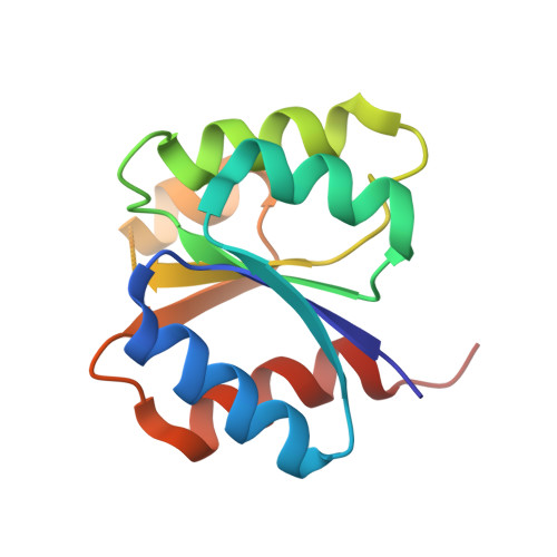
    DivK is an essential response regulator in the Gram-negative bacterium Caulobacter crescentus and functions in a complex phosphorelay system that precisely controls the sequence of developmental events during the cell division cycle. Structure determinations of this single domain response regulator at different pH values demonstrated that the five-stranded alpha/beta fold of the DivK protein is fully defined only at acidic pH. The crystal structures of the apoprotein and of metal-bound DivK complexes at higher pH values revealed a synergistic pH- and cation binding-induced flexibility of the beta4-alpha4 loop and of the alpha4 helix. This motion increases the solvent accessibility of the single cysteine residue in the protein. Solution state studies demonstrated a 200-fold pH-dependent increase in the affinity of manganese for the protein between pH 6.0 and 8.5 that seems to involve deprotonation of an acido-basic couple. Taken together, these results suggest that flexibility of critical regions of the protein, ionization of the cysteine 99 residue and improved K(D) values for the catalytic metal ion are coupled events. We propose that the molecular events observed in the isolated protein may be required for DivK activation and that they may be achieved in vivo through the specific protein-protein interactions between the response regulator and its cognate kinases.


  • Organizational Affiliation

    Groupe de Cristallographie Biologique, IPBS-CNRS, 205 route de Narbonne, 31077 Toulouse, France.


Macromolecules
Find similar proteins by:  (by identity cutoff)  |  3D Structure
Entity ID: 1
MoleculeChains Sequence LengthOrganismDetailsImage
cell division response regulator DivK124Caulobacter vibrioidesMutation(s): 0 
Gene Names: divk
UniProt
Find proteins for Q9A5I4 (Caulobacter vibrioides (strain ATCC 19089 / CB15))
Explore Q9A5I4 
Go to UniProtKB:  Q9A5I4
Entity Groups  
Sequence Clusters30% Identity50% Identity70% Identity90% Identity95% Identity100% Identity
UniProt GroupQ9A5I4
Sequence Annotations
Expand
  • Reference Sequence
Small Molecules
Ligands 1 Unique
IDChains Name / Formula / InChI Key2D Diagram3D Interactions
MG
Query on MG

Download Ideal Coordinates CCD File 
B [auth A]MAGNESIUM ION
Mg
JLVVSXFLKOJNIY-UHFFFAOYSA-N
Experimental Data & Validation

Experimental Data

  • Method: X-RAY DIFFRACTION
  • Resolution: 1.41 Å
  • R-Value Free: 0.215 
  • R-Value Work: 0.207 
  • Space Group: P 21 21 21
Unit Cell:
Length ( Å )Angle ( ˚ )
a = 36.77α = 90
b = 41.07β = 90
c = 67.12γ = 90
Software Package:
Software NamePurpose
MOSFLMdata reduction
SCALAdata scaling
CNSrefinement
CCP4data scaling
CNSphasing

Structure Validation

View Full Validation Report


Entry History 

Deposition Data

Revision History  (Full details and data files)

  • Version 1.0: 2002-12-04
    Type: Initial release
  • Version 1.1: 2008-04-28
    Changes: Version format compliance
  • Version 1.2: 2011-07-13
    Changes: Version format compliance
  • Version 1.3: 2017-02-01
    Changes: Database references, Structure summary
  • Version 1.4: 2024-02-14
    Changes: Data collection, Database references, Derived calculations
  • Version 1.5: 2024-04-03
    Changes: Refinement description