1MAI

STRUCTURE OF THE PLECKSTRIN HOMOLOGY DOMAIN FROM PHOSPHOLIPASE C DELTA IN COMPLEX WITH INOSITOL TRISPHOSPHATE


Experimental Data Snapshot

  • Method: X-RAY DIFFRACTION
  • Resolution: 1.90 Å
  • R-Value Free: 0.291 
  • R-Value Work: 0.205 
  • R-Value Observed: 0.205 

wwPDB Validation   3D Report Full Report


This is version 1.3 of the entry. See complete history


Literature

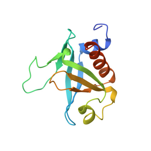
Structure of the high affinity complex of inositol trisphosphate with a phospholipase C pleckstrin homology domain.

Ferguson, K.M.Lemmon, M.A.Schlessinger, J.Sigler, P.B.

(1995) Cell 83: 1037-1046

  • DOI: https://doi.org/10.1016/0092-8674(95)90219-8
  • Primary Citation of Related Structures:  
    1MAI

  • PubMed Abstract: 

    The X-ray crystal structure of the high affinity complex between the pleckstrin homology (PH) domain from rat phospholipase C-delta 1 (PLC-delta 1) and inositol-(1,4,5)-trisphosphate (Ins(1,4,5)P3) has been refined to 1.9 A resolution. The domain fold is similar to others of known structure. Ins(1,4,5)P3 binds on the positively charged face of the electrostatically polarized domain, interacting predominantly with the beta 1/beta 2 and beta 3/beta 4 loops. The 4- and 5-phosphate groups of Ins(1,4,5)P3 interact much more extensively than the 1-phosphate. Two amino acids in the PLC-delta 1 PH domain that contact Ins(1,4,5)P3 have counterparts in the Bruton's tyrosine kinase (Btk) PH domain, where mutational changes cause inherited agammaglobulinemia, suggesting a mechanism for loss of function in Btk mutants. Using electrostatics and varying levels of head-group specificity, PH domains may localize and orient signaling proteins, providing a general membrane targeting and regulatory function.


  • Organizational Affiliation

    Department of Chemistry, Yale University, New Haven, Connecticut 06510, USA.


Macromolecules
Find similar proteins by:  (by identity cutoff)  |  3D Structure
Entity ID: 1
MoleculeChains Sequence LengthOrganismDetailsImage
PHOSPHOLIPASE C DELTA-1131Rattus norvegicusMutation(s): 0 
EC: 3.1.4.11
UniProt
Find proteins for P10688 (Rattus norvegicus)
Explore P10688 
Go to UniProtKB:  P10688
Entity Groups  
Sequence Clusters30% Identity50% Identity70% Identity90% Identity95% Identity100% Identity
UniProt GroupP10688
Sequence Annotations
Expand
  • Reference Sequence
Small Molecules
Ligands 1 Unique
IDChains Name / Formula / InChI Key2D Diagram3D Interactions
I3P
Query on I3P

Download Ideal Coordinates CCD File 
B [auth A]D-MYO-INOSITOL-1,4,5-TRIPHOSPHATE
C6 H15 O15 P3
MMWCIQZXVOZEGG-XJTPDSDZSA-N
Binding Affinity Annotations 
IDSourceBinding Affinity
I3P Binding MOAD:  1MAI Kd: 210 (nM) from 1 assay(s)
PDBBind:  1MAI Kd: 210 (nM) from 1 assay(s)
Experimental Data & Validation

Experimental Data

  • Method: X-RAY DIFFRACTION
  • Resolution: 1.90 Å
  • R-Value Free: 0.291 
  • R-Value Work: 0.205 
  • R-Value Observed: 0.205 
  • Space Group: P 43 21 2
Unit Cell:
Length ( Å )Angle ( ˚ )
a = 59.4α = 90
b = 59.4β = 90
c = 80.7γ = 90
Software Package:
Software NamePurpose
X-PLORmodel building
X-PLORrefinement
DENZOdata reduction
X-PLORphasing

Structure Validation

View Full Validation Report


Entry History 

Deposition Data

Revision History  (Full details and data files)

  • Version 1.0: 1996-11-08
    Type: Initial release
  • Version 1.1: 2008-03-24
    Changes: Version format compliance
  • Version 1.2: 2011-07-13
    Changes: Version format compliance
  • Version 1.3: 2024-02-14
    Changes: Data collection, Database references, Derived calculations, Other