1M74

Crystal structure of Mg-ADP-bound SecA from Bacillus subtilis


Experimental Data Snapshot

  • Method: X-RAY DIFFRACTION
  • Resolution: 3.00 Å
  • R-Value Free: 0.291 
  • R-Value Work: 0.217 
  • R-Value Observed: 0.217 

wwPDB Validation   3D Report Full Report


Ligand Structure Quality Assessment 


This is version 1.3 of the entry. See complete history


Literature

Nucleotide Control of Interdomain Interactions in the Conformational Reaction Cycle of SecA

Hunt, J.F.Weinkauf, S.Henry, L.Fak, J.J.McNicholas, P.Oliver, D.B.Deisenhofer, J.

(2002) Science 297: 2018-2026

  • DOI: https://doi.org/10.1126/science.1074424
  • Primary Citation of Related Structures:  
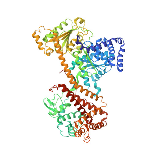
    1M6N, 1M74

  • PubMed Abstract: 

    The SecA adenosine triphosphatase (ATPase) mediates extrusion of the amino termini of secreted proteins from the eubacterial cytosol based on cycles of reversible binding to the SecYEG translocon. We have determined the crystal structure of SecA with and without magnesium-adenosine diphosphate bound to the high-affinity ATPase site at 3.0 and 2.7 angstrom resolution, respectively. Candidate sites for preprotein binding are located on a surface containing the SecA epitopes exposed to the periplasm upon binding to SecYEG and are thus positioned to deliver preprotein to SecYEG. Comparisons with structurally related ATPases, including superfamily I and II ATP-dependent helicases, suggest that the interaction geometry of the tandem motor domains in SecA is modulated by nucleotide binding, which is shown by fluorescence anisotropy experiments to reverse an endothermic domain-dissociation reaction hypothesized to gate binding to SecYEG.


  • Organizational Affiliation

    Department of Biological Sciences, 702A Fairchild Center, MC2434, Columbia University, New York, NY 10027, USA. hunt@sid.bio.columbia.edu


Macromolecules
Find similar proteins by:  (by identity cutoff)  |  3D Structure
Entity ID: 1
MoleculeChains Sequence LengthOrganismDetailsImage
Preprotein translocase secA802Bacillus subtilisMutation(s): 0 
Gene Names: Div
UniProt
Find proteins for P28366 (Bacillus subtilis (strain 168))
Explore P28366 
Go to UniProtKB:  P28366
Entity Groups  
Sequence Clusters30% Identity50% Identity70% Identity90% Identity95% Identity100% Identity
UniProt GroupP28366
Sequence Annotations
Expand
  • Reference Sequence
Small Molecules
Experimental Data & Validation

Experimental Data

  • Method: X-RAY DIFFRACTION
  • Resolution: 3.00 Å
  • R-Value Free: 0.291 
  • R-Value Work: 0.217 
  • R-Value Observed: 0.217 
  • Space Group: P 31 1 2
Unit Cell:
Length ( Å )Angle ( ˚ )
a = 131.245α = 90
b = 131.245β = 90
c = 150.453γ = 120
Software Package:
Software NamePurpose
X-PLORrefinement
DENZOdata reduction
SCALEPACKdata scaling

Structure Validation

View Full Validation Report


Ligand Structure Quality Assessment 


Entry History 

Deposition Data

Revision History  (Full details and data files)

  • Version 1.0: 2002-09-20
    Type: Initial release
  • Version 1.1: 2008-04-28
    Changes: Version format compliance
  • Version 1.2: 2011-07-13
    Changes: Derived calculations, Version format compliance
  • Version 1.3: 2024-02-14
    Changes: Data collection, Database references, Derived calculations, Refinement description